Mz Faizah, Y Kanaheswari, Cr Thambidorai, Ma Zulfiqar
{"title":"Echocontrast cystosonography versus micturating cystourethrography in the detection of vesicoureteric reflux.","authors":"Mz Faizah, Y Kanaheswari, Cr Thambidorai, Ma Zulfiqar","doi":"10.2349/biij.7.1.e7","DOIUrl":null,"url":null,"abstract":"<p><strong>Purpose: </strong>To compare echocontrast cystosonography (ECS) using in-vivo agitated saline with fluoroscopic micturating cystourethrography (MCU) in the detection and grading of vesicoureteric reflux (VUR).</p><p><strong>Materials and methods: </strong>This was a prospective study of 25 children, who had MCU between 2007 and 2009. ECS was performed and findings documented prior to MCU. Baseline renal and bladder sonograms were obtained. The bladder was filled with normal saline followed by introduction of 10-20 mls of air to generate microbubbles. Detection of VUR was based on two sonographic criteria: (1) presence of microbubbles in the pelvicaliceal system (PCS), and (2) increase in dilatation of the PCS. VUR was graded as (1) Grade I: microbubbles seen in ureter only; (2) Grade II: microbubbles seen in non-dilated PCS; and (3) Grade III-V: microbubbles seen in dilated PCS. The ECS findings were compared using MCU as the gold standard.</p><p><strong>Results: </strong>Of the 50 kidney-ureter (K-U) units studied, ECS detected 9 of 10 K-U units with VUR on MCU. ECS did not detect a Grade II VUR. The sensitivity, specificity, accuracy, positive predictive value and negative predictive value for criterion 1 was 90%, 87.5%, 88%, 64.3% and 97%, respectively, compared to criterion 2 which was 70%, 90%, 86%, 64% and 92%, respectively. The grading of VUR was similar on both ECS and MCU except for one case.</p><p><strong>Conclusion: </strong>ECS using agitated saline was a sensitive technique for the detection of VUR. ECS grading was comparable with MCU grading of VUR.</p>","PeriodicalId":89331,"journal":{"name":"Biomedical imaging and intervention journal","volume":"7 1","pages":"e7"},"PeriodicalIF":0.0000,"publicationDate":"2011-01-01","publicationTypes":"Journal Article","fieldsOfStudy":null,"isOpenAccess":false,"openAccessPdf":"https://sci-hub-pdf.com/10.2349/biij.7.1.e7","citationCount":"6","resultStr":null,"platform":"Semanticscholar","paperid":null,"PeriodicalName":"Biomedical imaging and intervention journal","FirstCategoryId":"1085","ListUrlMain":"https://doi.org/10.2349/biij.7.1.e7","RegionNum":0,"RegionCategory":null,"ArticlePicture":[],"TitleCN":null,"AbstractTextCN":null,"PMCID":null,"EPubDate":"","PubModel":"","JCR":"","JCRName":"","Score":null,"Total":0}
引用次数: 6
Abstract
Purpose: To compare echocontrast cystosonography (ECS) using in-vivo agitated saline with fluoroscopic micturating cystourethrography (MCU) in the detection and grading of vesicoureteric reflux (VUR).
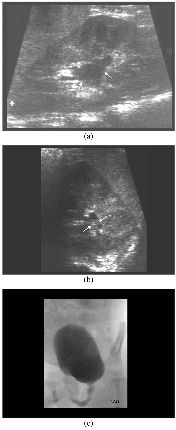
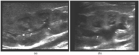
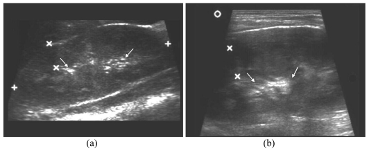
Materials and methods: This was a prospective study of 25 children, who had MCU between 2007 and 2009. ECS was performed and findings documented prior to MCU. Baseline renal and bladder sonograms were obtained. The bladder was filled with normal saline followed by introduction of 10-20 mls of air to generate microbubbles. Detection of VUR was based on two sonographic criteria: (1) presence of microbubbles in the pelvicaliceal system (PCS), and (2) increase in dilatation of the PCS. VUR was graded as (1) Grade I: microbubbles seen in ureter only; (2) Grade II: microbubbles seen in non-dilated PCS; and (3) Grade III-V: microbubbles seen in dilated PCS. The ECS findings were compared using MCU as the gold standard.
Results: Of the 50 kidney-ureter (K-U) units studied, ECS detected 9 of 10 K-U units with VUR on MCU. ECS did not detect a Grade II VUR. The sensitivity, specificity, accuracy, positive predictive value and negative predictive value for criterion 1 was 90%, 87.5%, 88%, 64.3% and 97%, respectively, compared to criterion 2 which was 70%, 90%, 86%, 64% and 92%, respectively. The grading of VUR was similar on both ECS and MCU except for one case.
Conclusion: ECS using agitated saline was a sensitive technique for the detection of VUR. ECS grading was comparable with MCU grading of VUR.

 求助内容:
求助内容: 应助结果提醒方式:


