{"title":"Classification and diagnosis of ear malformations.","authors":"Sylva Bartel-Friedrich, Cornelia Wulke","doi":"","DOIUrl":null,"url":null,"abstract":"<p><p>In the ENT region 50% of the malformations affect the ear. Malformations of the outer and middle ear are predominantly unilateral (ca. 70-90%) and mostly involve the right ear. Inner ear malformations can be unilateral or bilateral. The incidence of ear malformations is approximately 1 in 3800 newborns. Ear malformations may be genetic (associated with syndromes or not, with family history, spontaneous mutations) or acquired in nature. Malformations can affect the outer ear (pinna and external auditory canal, EAC), middle ear and inner ear, not infrequently in combination. Formal classification is advisable in order to be able to predict the prognosis and compare treatment schedules. Various classifications have been proposed: pinna and EAC malformations according to Weerda [1], middle ear malformations according to Kösling [2], and inner ear malformations according to Jackler [3], [4], to Marangos [5] and to Sennaroglu [6]. Additionally, we describe Altmann's classification of atresia auris congenita [7] and the Siegert-Mayer-Weerda score [8] for EAC and middle ear malformations, systems of great practicability that are in widespread clinical use. The diagnostic steps include clinical examination, audiological testing, genetic analysis and, especially, CT and MRI. These imaging methods are most usefully employed in combination. Precise description of the malformations by means of CT and MRI is indispensable for the planning and successful outcome of operative ear reconstruction and rehabilitation procedures, including cochlear implantation.</p>","PeriodicalId":89377,"journal":{"name":"GMS current topics in otorhinolaryngology, head and neck surgery","volume":"6 ","pages":"Doc05"},"PeriodicalIF":0.0000,"publicationDate":"2007-01-01","publicationTypes":"Journal Article","fieldsOfStudy":null,"isOpenAccess":false,"openAccessPdf":"https://ftp.ncbi.nlm.nih.gov/pub/pmc/oa_pdf/d7/eb/CTO-06-05.PMC3199848.pdf","citationCount":"0","resultStr":null,"platform":"Semanticscholar","paperid":null,"PeriodicalName":"GMS current topics in otorhinolaryngology, head and neck surgery","FirstCategoryId":"1085","ListUrlMain":"","RegionNum":0,"RegionCategory":null,"ArticlePicture":[],"TitleCN":null,"AbstractTextCN":null,"PMCID":null,"EPubDate":"2008/3/14 0:00:00","PubModel":"Epub","JCR":"","JCRName":"","Score":null,"Total":0}
引用次数: 0
Abstract
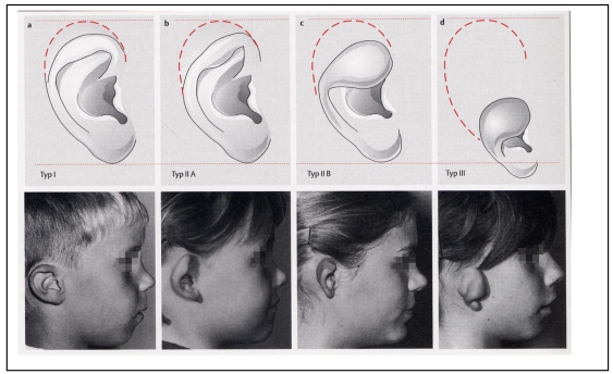
In the ENT region 50% of the malformations affect the ear. Malformations of the outer and middle ear are predominantly unilateral (ca. 70-90%) and mostly involve the right ear. Inner ear malformations can be unilateral or bilateral. The incidence of ear malformations is approximately 1 in 3800 newborns. Ear malformations may be genetic (associated with syndromes or not, with family history, spontaneous mutations) or acquired in nature. Malformations can affect the outer ear (pinna and external auditory canal, EAC), middle ear and inner ear, not infrequently in combination. Formal classification is advisable in order to be able to predict the prognosis and compare treatment schedules. Various classifications have been proposed: pinna and EAC malformations according to Weerda [1], middle ear malformations according to Kösling [2], and inner ear malformations according to Jackler [3], [4], to Marangos [5] and to Sennaroglu [6]. Additionally, we describe Altmann's classification of atresia auris congenita [7] and the Siegert-Mayer-Weerda score [8] for EAC and middle ear malformations, systems of great practicability that are in widespread clinical use. The diagnostic steps include clinical examination, audiological testing, genetic analysis and, especially, CT and MRI. These imaging methods are most usefully employed in combination. Precise description of the malformations by means of CT and MRI is indispensable for the planning and successful outcome of operative ear reconstruction and rehabilitation procedures, including cochlear implantation.

 求助内容:
求助内容: 应助结果提醒方式:


