Frederico F R Maia, Patrícia S Matos, Bradley P Silva, Ana T Pallone, Elizabeth J Pavin, José Vassallo, Denise E Zantut-Wittmann
{"title":"Role of ultrasound, clinical and scintigraphyc parameters to predict malignancy in thyroid nodule.","authors":"Frederico F R Maia, Patrícia S Matos, Bradley P Silva, Ana T Pallone, Elizabeth J Pavin, José Vassallo, Denise E Zantut-Wittmann","doi":"10.1186/1758-3284-3-17","DOIUrl":null,"url":null,"abstract":"<p><strong>Background: </strong>This study aimed to evaluate clinical, laboratory, ultrasound (US) and scintigraphyc parameters in thyroid nodule and to develop an auxiliary model for clinical application in the diagnosis of malignancy.</p><p><strong>Methods: </strong>We assessed 143 patients who were surgically treated at a single center, 65% (93) benign vs. 35% (50) malignant lesions at final histology (1998-2008). The clinical, laboratory, scintigraphyc and US features were compared and a prediction model was designed after the multivariate analysis.</p><p><strong>Results: </strong>There were no differences in gender, serum TSH and FT4 levels, thyroid auto-antibodies (TAb), thyroid dysfunction and scintigraphyc results (P=0.33) between benign and malignant nodule groups. The sonographic study showed differences when the presence of suspected characteristics was found in the nodules of the malignant lesions group, such as: microcalcifications, central flow, border irregularity and hypoechogenicity. After the multivariate analysis the model obtained showed age (>39 years), border irregularity, microcalcifications and nodule size over 2 cm as predictive factors of malignancy, featuring 81.7% of accuracy.</p><p><strong>Conclusions: </strong>This study confirmed a significant increase of risk for malignancy in patients of over 39 years and with suspicious features at US.</p>","PeriodicalId":49195,"journal":{"name":"Head and Neck Optical Diagnostics Society","volume":"3 ","pages":"17"},"PeriodicalIF":0.0000,"publicationDate":"2011-03-22","publicationTypes":"Journal Article","fieldsOfStudy":null,"isOpenAccess":false,"openAccessPdf":"https://sci-hub-pdf.com/10.1186/1758-3284-3-17","citationCount":"35","resultStr":null,"platform":"Semanticscholar","paperid":null,"PeriodicalName":"Head and Neck Optical Diagnostics Society","FirstCategoryId":"1085","ListUrlMain":"https://doi.org/10.1186/1758-3284-3-17","RegionNum":0,"RegionCategory":null,"ArticlePicture":[],"TitleCN":null,"AbstractTextCN":null,"PMCID":null,"EPubDate":"","PubModel":"","JCR":"","JCRName":"","Score":null,"Total":0}
引用次数: 35
Abstract
Background: This study aimed to evaluate clinical, laboratory, ultrasound (US) and scintigraphyc parameters in thyroid nodule and to develop an auxiliary model for clinical application in the diagnosis of malignancy.
Methods: We assessed 143 patients who were surgically treated at a single center, 65% (93) benign vs. 35% (50) malignant lesions at final histology (1998-2008). The clinical, laboratory, scintigraphyc and US features were compared and a prediction model was designed after the multivariate analysis.
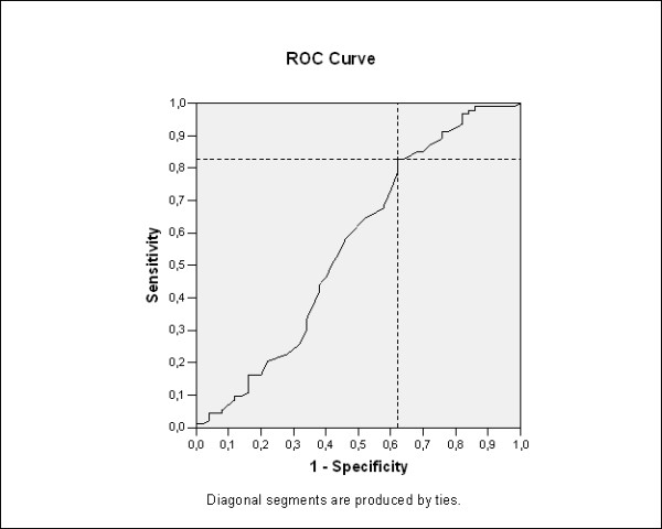
Results: There were no differences in gender, serum TSH and FT4 levels, thyroid auto-antibodies (TAb), thyroid dysfunction and scintigraphyc results (P=0.33) between benign and malignant nodule groups. The sonographic study showed differences when the presence of suspected characteristics was found in the nodules of the malignant lesions group, such as: microcalcifications, central flow, border irregularity and hypoechogenicity. After the multivariate analysis the model obtained showed age (>39 years), border irregularity, microcalcifications and nodule size over 2 cm as predictive factors of malignancy, featuring 81.7% of accuracy.
Conclusions: This study confirmed a significant increase of risk for malignancy in patients of over 39 years and with suspicious features at US.
 求助内容:
求助内容: 应助结果提醒方式:


