Mutual information identifies sequence positions conserved within the nuclear receptor superfamily: approach reveals functionally important regions for DNA binding specificity.
{"title":"Mutual information identifies sequence positions conserved within the nuclear receptor superfamily: approach reveals functionally important regions for DNA binding specificity.","authors":"Scooter Willis, Patrick R Griffin","doi":"10.1621/nrs.09001","DOIUrl":null,"url":null,"abstract":"<p><p>Members of the nuclear receptor superfamily differentiate in terms of specificity for DNA recognition and binding, oligomeric state, and ligand binding. The wide range of specificities are impressive given the high degree of sequence conservation in the DNA binding domain (DBD) and moderate sequence conservation with high structural similarity within the ligand binding domains (LBDs). Determining sequence positions that are conserved within nuclear receptor subfamilies can provide important indicators into the structural dynamics that translate to oligomeric state of the active receptor, DNA binding specificity and ligand affinity and selectivity. Here we present a method to analyze sequence data from all nuclear receptors that facilitates detection of co-evolving pairs using Mutual Information (MI). Using this method we demonstrate that MI can reveal functionally important sequence positions within the superfamily and the approach identified three sequence positions that have conserved sequence patterns across all nuclear receptors and subfamilies. Interestingly, two of the sequence positions identified are located within the DBD CII and the third was within Helix c of the DBD. These sequences are located within the heterodimer interface of PPARγ (CII) and RXRα (Helix c) based on PDB:3DZU. Helix c of PPARγ, which is not involved in the DBD dimer interface, binds the minor groove in the 5' flanking region in a consensus PPARγ response element (PPRE) and the corresponding RXRα (CII) is found in the 3' flanking region of RXRE (3DZU). As these three sequence positions represent unique identifiers for all nuclear receptors and they are located within the dimer interface of PPARγ-RXRα DBD (3DZU) interfacing with the flanking regions of the NRRE, we conclude they are critical sequence positions perhaps dictating nuclear receptor (NR) DNA binding specificity.</p>","PeriodicalId":87415,"journal":{"name":"Nuclear receptor signaling","volume":"9 ","pages":"e001"},"PeriodicalIF":0.0000,"publicationDate":"2011-02-25","publicationTypes":"Journal Article","fieldsOfStudy":null,"isOpenAccess":false,"openAccessPdf":"https://sci-hub-pdf.com/10.1621/nrs.09001","citationCount":"5","resultStr":null,"platform":"Semanticscholar","paperid":null,"PeriodicalName":"Nuclear receptor signaling","FirstCategoryId":"1085","ListUrlMain":"https://doi.org/10.1621/nrs.09001","RegionNum":0,"RegionCategory":null,"ArticlePicture":[],"TitleCN":null,"AbstractTextCN":null,"PMCID":null,"EPubDate":"","PubModel":"","JCR":"","JCRName":"","Score":null,"Total":0}
引用次数: 5
Abstract
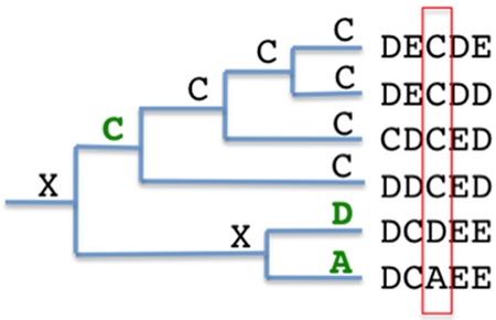
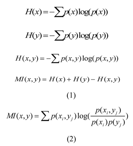
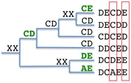
Members of the nuclear receptor superfamily differentiate in terms of specificity for DNA recognition and binding, oligomeric state, and ligand binding. The wide range of specificities are impressive given the high degree of sequence conservation in the DNA binding domain (DBD) and moderate sequence conservation with high structural similarity within the ligand binding domains (LBDs). Determining sequence positions that are conserved within nuclear receptor subfamilies can provide important indicators into the structural dynamics that translate to oligomeric state of the active receptor, DNA binding specificity and ligand affinity and selectivity. Here we present a method to analyze sequence data from all nuclear receptors that facilitates detection of co-evolving pairs using Mutual Information (MI). Using this method we demonstrate that MI can reveal functionally important sequence positions within the superfamily and the approach identified three sequence positions that have conserved sequence patterns across all nuclear receptors and subfamilies. Interestingly, two of the sequence positions identified are located within the DBD CII and the third was within Helix c of the DBD. These sequences are located within the heterodimer interface of PPARγ (CII) and RXRα (Helix c) based on PDB:3DZU. Helix c of PPARγ, which is not involved in the DBD dimer interface, binds the minor groove in the 5' flanking region in a consensus PPARγ response element (PPRE) and the corresponding RXRα (CII) is found in the 3' flanking region of RXRE (3DZU). As these three sequence positions represent unique identifiers for all nuclear receptors and they are located within the dimer interface of PPARγ-RXRα DBD (3DZU) interfacing with the flanking regions of the NRRE, we conclude they are critical sequence positions perhaps dictating nuclear receptor (NR) DNA binding specificity.

 求助内容:
求助内容: 应助结果提醒方式:


