Ben Corry, Annette C Hurst, Prithwish Pal, Takeshi Nomura, Paul Rigby, Boris Martinac
{"title":"An improved open-channel structure of MscL determined from FRET confocal microscopy and simulation.","authors":"Ben Corry, Annette C Hurst, Prithwish Pal, Takeshi Nomura, Paul Rigby, Boris Martinac","doi":"10.1085/jgp.200910376","DOIUrl":null,"url":null,"abstract":"<p><p>Mechanosensitive channels act as molecular transducers of mechanical force exerted on the membrane of living cells by opening in response to membrane bilayer deformations occurring in physiological processes such as touch, hearing, blood pressure regulation, and osmoregulation. Here, we determine the likely structure of the open state of the mechanosensitive channel of large conductance using a combination of patch clamp, fluorescence resonance energy transfer (FRET) spectroscopy, data from previous electron paramagnetic resonance experiments, and molecular and Brownian dynamics simulations. We show that structural rearrangements of the protein can be measured in similar conditions as patch clamp recordings while controlling the state of the pore in its natural lipid environment by modifying the lateral pressure distribution via the lipid bilayer. Transition to the open state is less dramatic than previously proposed, while the N terminus remains anchored at the surface of the membrane where it can either guide the tilt of or directly translate membrane tension to the conformation of the pore-lining helix. Combining FRET data obtained in physiological conditions with simulations is likely to be of great value for studying conformational changes in a range of multimeric membrane proteins.</p>","PeriodicalId":173753,"journal":{"name":"The Journal of General Physiology","volume":" ","pages":"483-94"},"PeriodicalIF":0.0000,"publicationDate":"2010-10-01","publicationTypes":"Journal Article","fieldsOfStudy":null,"isOpenAccess":false,"openAccessPdf":"https://sci-hub-pdf.com/10.1085/jgp.200910376","citationCount":"82","resultStr":null,"platform":"Semanticscholar","paperid":null,"PeriodicalName":"The Journal of General Physiology","FirstCategoryId":"3","ListUrlMain":"https://doi.org/10.1085/jgp.200910376","RegionNum":0,"RegionCategory":null,"ArticlePicture":[],"TitleCN":null,"AbstractTextCN":null,"PMCID":null,"EPubDate":"","PubModel":"","JCR":"","JCRName":"","Score":null,"Total":0}
引用次数: 82
Abstract
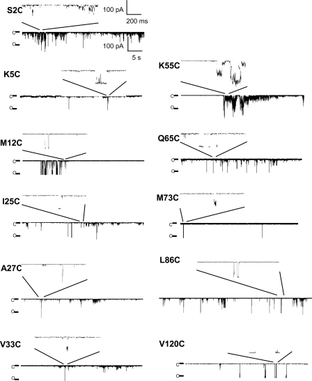
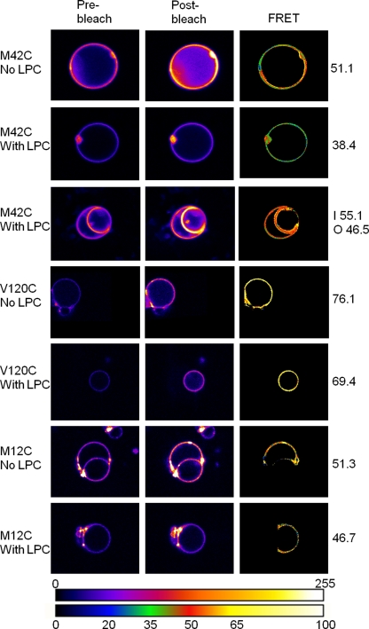
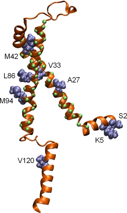
Mechanosensitive channels act as molecular transducers of mechanical force exerted on the membrane of living cells by opening in response to membrane bilayer deformations occurring in physiological processes such as touch, hearing, blood pressure regulation, and osmoregulation. Here, we determine the likely structure of the open state of the mechanosensitive channel of large conductance using a combination of patch clamp, fluorescence resonance energy transfer (FRET) spectroscopy, data from previous electron paramagnetic resonance experiments, and molecular and Brownian dynamics simulations. We show that structural rearrangements of the protein can be measured in similar conditions as patch clamp recordings while controlling the state of the pore in its natural lipid environment by modifying the lateral pressure distribution via the lipid bilayer. Transition to the open state is less dramatic than previously proposed, while the N terminus remains anchored at the surface of the membrane where it can either guide the tilt of or directly translate membrane tension to the conformation of the pore-lining helix. Combining FRET data obtained in physiological conditions with simulations is likely to be of great value for studying conformational changes in a range of multimeric membrane proteins.

 求助内容:
求助内容: 应助结果提醒方式:


