Endoscopic Ultrasound-Guided Fine Needle Aspiration (EUS-FNA) Diagnosis of Recurrent Anal Cancer After Chemoradiation and Negative Forceps Biopsies: A Case Report.
{"title":"Endoscopic Ultrasound-Guided Fine Needle Aspiration (EUS-FNA) Diagnosis of Recurrent Anal Cancer After Chemoradiation and Negative Forceps Biopsies: A Case Report.","authors":"Julia Leblanc, Pradermchai Kongkam","doi":"10.4137/cmo.s993","DOIUrl":null,"url":null,"abstract":"<p><p>A 69-year-old woman with a history of uT2 N0 post-treated anal squamous cell cancer (SCC) presented for EUS for perianal pain. Two months prior, a digital rectal examination was significant for an indurated lesion on the left lateral rectal wall just proximal to the dentate line. A sigmoidoscopy revealed mild narrowing of the anal canal and an ulcerated friable mucosa in the same area. A biopsy demonstrated ulceration without malignancy. EUS showed a hypoechoic, non-circumferential, left-sided distal rectal mass. EUS-FNA was performed. Cytology demonstrated poorly differentiated SCC. This was confirmed by subsequent surgical resection. While endoscopic biopsy of suspected anal recurrences is usually sufficient, histologic or cytologic confirmation are necessary, as radiation-induced changes are difficult to differentiate from tumor recurrence. This case demonstrates that EUS-FNA is useful in surveillance of anal SCC when there is a high clinical suspicion of recurrence.</p>","PeriodicalId":88451,"journal":{"name":"Clinical medicine. Oncology","volume":"3 ","pages":"59-62"},"PeriodicalIF":0.0000,"publicationDate":"2009-04-28","publicationTypes":"Journal Article","fieldsOfStudy":null,"isOpenAccess":false,"openAccessPdf":"https://sci-hub-pdf.com/10.4137/cmo.s993","citationCount":"3","resultStr":null,"platform":"Semanticscholar","paperid":null,"PeriodicalName":"Clinical medicine. Oncology","FirstCategoryId":"1085","ListUrlMain":"https://doi.org/10.4137/cmo.s993","RegionNum":0,"RegionCategory":null,"ArticlePicture":[],"TitleCN":null,"AbstractTextCN":null,"PMCID":null,"EPubDate":"","PubModel":"","JCR":"","JCRName":"","Score":null,"Total":0}
引用次数: 3
Abstract
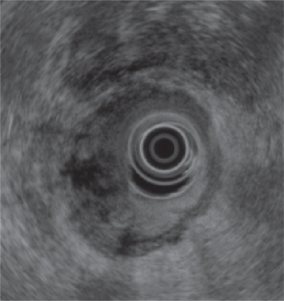
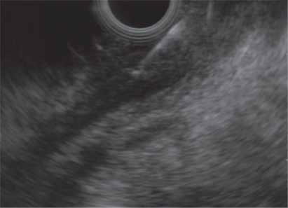
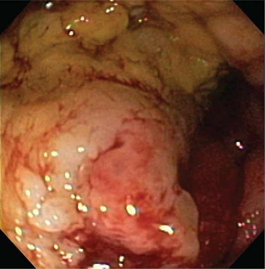
A 69-year-old woman with a history of uT2 N0 post-treated anal squamous cell cancer (SCC) presented for EUS for perianal pain. Two months prior, a digital rectal examination was significant for an indurated lesion on the left lateral rectal wall just proximal to the dentate line. A sigmoidoscopy revealed mild narrowing of the anal canal and an ulcerated friable mucosa in the same area. A biopsy demonstrated ulceration without malignancy. EUS showed a hypoechoic, non-circumferential, left-sided distal rectal mass. EUS-FNA was performed. Cytology demonstrated poorly differentiated SCC. This was confirmed by subsequent surgical resection. While endoscopic biopsy of suspected anal recurrences is usually sufficient, histologic or cytologic confirmation are necessary, as radiation-induced changes are difficult to differentiate from tumor recurrence. This case demonstrates that EUS-FNA is useful in surveillance of anal SCC when there is a high clinical suspicion of recurrence.

 求助内容:
求助内容: 应助结果提醒方式:


