Eli J Weinberg, Peter J Mack, Frederick J Schoen, Guillermo García-Cardeña, Mohammad R Kaazempur Mofrad
{"title":"Hemodynamic environments from opposing sides of human aortic valve leaflets evoke distinct endothelial phenotypes in vitro.","authors":"Eli J Weinberg, Peter J Mack, Frederick J Schoen, Guillermo García-Cardeña, Mohammad R Kaazempur Mofrad","doi":"10.1007/s10558-009-9089-9","DOIUrl":null,"url":null,"abstract":"<p><p>The regulation of valvular endothelial phenotypes by the hemodynamic environments of the human aortic valve is poorly understood. The nodular lesions of calcific aortic stenosis (CAS) develop predominantly beneath the aortic surface of the valve leaflets in the valvular fibrosa layer. However, the mechanisms of this regional localization remain poorly characterized. In this study, we combine numerical simulation with in vitro experimentation to investigate the hypothesis that the previously documented differences between valve endothelial phenotypes are linked to distinct hemodynamic environments characteristic of these individual anatomical locations. A finite-element model of the aortic valve was created, describing the dynamic motion of the valve cusps and blood in the valve throughout the cardiac cycle. A fluid mesh with high resolution on the fluid boundary was used to allow accurate computation of the wall shear stresses. This model was used to compute two distinct shear stress waveforms, one for the ventricular surface and one for the aortic surface. These waveforms were then applied experimentally to cultured human endothelial cells and the expression of several pathophysiological relevant genes was assessed. Compared to endothelial cells subjected to shear stress waveforms representative of the aortic face, the endothelial cells subjected to the ventricular waveform showed significantly increased expression of the \"atheroprotective\" transcription factor Kruppel-like factor 2 (KLF2) and the matricellular protein Nephroblastoma overexpressed (NOV), and suppressed expression of chemokine Monocyte-chemotactic protein-1 (MCP-1). Our observations suggest that the difference in shear stress waveforms between the two sides of the aortic valve leaflet may contribute to the documented differential side-specific gene expression, and may be relevant for the development and progression of CAS and the potential role of endothelial mechanotransduction in this disease.</p>","PeriodicalId":55275,"journal":{"name":"Cardiovascular Engineering (dordrecht, Netherlands)","volume":"10 1","pages":"5-11"},"PeriodicalIF":0.0000,"publicationDate":"2010-03-01","publicationTypes":"Journal Article","fieldsOfStudy":null,"isOpenAccess":false,"openAccessPdf":"https://sci-hub-pdf.com/10.1007/s10558-009-9089-9","citationCount":"63","resultStr":null,"platform":"Semanticscholar","paperid":null,"PeriodicalName":"Cardiovascular Engineering (dordrecht, Netherlands)","FirstCategoryId":"1085","ListUrlMain":"https://doi.org/10.1007/s10558-009-9089-9","RegionNum":0,"RegionCategory":null,"ArticlePicture":[],"TitleCN":null,"AbstractTextCN":null,"PMCID":null,"EPubDate":"","PubModel":"","JCR":"","JCRName":"","Score":null,"Total":0}
引用次数: 63
Abstract
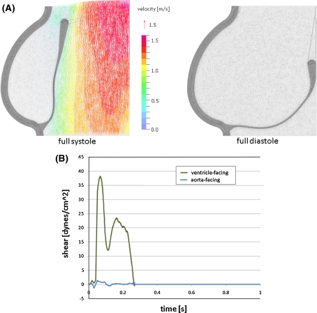
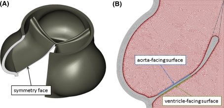
The regulation of valvular endothelial phenotypes by the hemodynamic environments of the human aortic valve is poorly understood. The nodular lesions of calcific aortic stenosis (CAS) develop predominantly beneath the aortic surface of the valve leaflets in the valvular fibrosa layer. However, the mechanisms of this regional localization remain poorly characterized. In this study, we combine numerical simulation with in vitro experimentation to investigate the hypothesis that the previously documented differences between valve endothelial phenotypes are linked to distinct hemodynamic environments characteristic of these individual anatomical locations. A finite-element model of the aortic valve was created, describing the dynamic motion of the valve cusps and blood in the valve throughout the cardiac cycle. A fluid mesh with high resolution on the fluid boundary was used to allow accurate computation of the wall shear stresses. This model was used to compute two distinct shear stress waveforms, one for the ventricular surface and one for the aortic surface. These waveforms were then applied experimentally to cultured human endothelial cells and the expression of several pathophysiological relevant genes was assessed. Compared to endothelial cells subjected to shear stress waveforms representative of the aortic face, the endothelial cells subjected to the ventricular waveform showed significantly increased expression of the "atheroprotective" transcription factor Kruppel-like factor 2 (KLF2) and the matricellular protein Nephroblastoma overexpressed (NOV), and suppressed expression of chemokine Monocyte-chemotactic protein-1 (MCP-1). Our observations suggest that the difference in shear stress waveforms between the two sides of the aortic valve leaflet may contribute to the documented differential side-specific gene expression, and may be relevant for the development and progression of CAS and the potential role of endothelial mechanotransduction in this disease.

 求助内容:
求助内容: 应助结果提醒方式:


