John C Licciardone, Kimberly G Fulda, Scott T Stoll, Russell G Gamber, A Clifton Cage
{"title":"A case-control study of osteopathic palpatory findings in type 2 diabetes mellitus.","authors":"John C Licciardone, Kimberly G Fulda, Scott T Stoll, Russell G Gamber, A Clifton Cage","doi":"10.1186/1750-4732-1-6","DOIUrl":null,"url":null,"abstract":"<p><strong>Background: </strong>Although type 2 diabetes mellitus is often managed by osteopathic physicians, osteopathic palpatory findings in this disease have not been adequately studied.</p><p><strong>Methods: </strong>A case-control study was used to measure the association between type 2 diabetes mellitus and a series of 30 osteopathic palpatory findings. The latter included skin changes, trophic changes, tissue changes, tenderness, and immobility at spinal segmental levels T5-T7, T8-T10, and T11-L2 bilaterally. Logistic regression models that adjusted for age, sex, and comorbid conditions were used to compute odds ratios (ORs) and 95% confidence intervals (CIs) for the associations between type 2 diabetes mellitus and each of these findings.</p><p><strong>Results and discussion: </strong>A total of 92 subjects were included in the study. After controlling for age, sex, hypertension, and clinical depression, the only significant finding was an association between type 2 diabetes mellitus and tissue changes at T11-L2 on the right side (OR, 5.54; 95% CI, 1.76-17.47; P = .003). Subgroup analyses of subjects with type 2 diabetes mellitus and hypertension demonstrated significant associations with tissue changes at T11-L2 bilaterally (OR, 27.38; 95% CI, 1.75-428; P = .02 for the left side and OR, 24.00; 95% CI, 1.51-382; P = .02 for the right side). Among subjects with type 2 diabetes mellitus and hypertension, there was also a strong diabetes mellitus duration effect for tissue changes at T11-L2 bilaterally (OR, 12.00; 95% CI, 1.02-141; P = .05 for short duration vs. OR, 32.00; 95% CI, 2.29-448; P = .01 for long duration on the left side; and OR, 17.33; 95% CI, 1.39-217; P = .03 for short duration vs. OR, 32.00; 95% CI, 2.29-448; P = .01 for long duration on the right side).</p><p><strong>Conclusion: </strong>The only consistent finding in this study was an association between type 2 diabetes mellitus and tissue changes at T11-L2 on the right side. Potential explanations for this finding include reflex viscerosomatic changes directly related to the progression of type 2 diabetes mellitus, a spurious association attributable to confounding visceral diseases, or a chance observation unrelated to type 2 diabetes mellitus. Larger prospective studies are needed to better study osteopathic palpatory findings in type 2 diabetes mellitus.</p>","PeriodicalId":87450,"journal":{"name":"Osteopathic medicine and primary care","volume":null,"pages":null},"PeriodicalIF":0.0000,"publicationDate":"2007-02-08","publicationTypes":"Journal Article","fieldsOfStudy":null,"isOpenAccess":false,"openAccessPdf":"https://sci-hub-pdf.com/10.1186/1750-4732-1-6","citationCount":"1","resultStr":null,"platform":"Semanticscholar","paperid":null,"PeriodicalName":"Osteopathic medicine and primary care","FirstCategoryId":"1085","ListUrlMain":"https://doi.org/10.1186/1750-4732-1-6","RegionNum":0,"RegionCategory":null,"ArticlePicture":[],"TitleCN":null,"AbstractTextCN":null,"PMCID":null,"EPubDate":"","PubModel":"","JCR":"","JCRName":"","Score":null,"Total":0}
引用次数: 1
Abstract
Background: Although type 2 diabetes mellitus is often managed by osteopathic physicians, osteopathic palpatory findings in this disease have not been adequately studied.
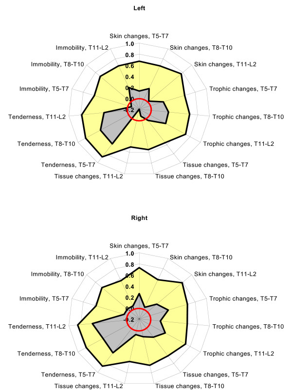
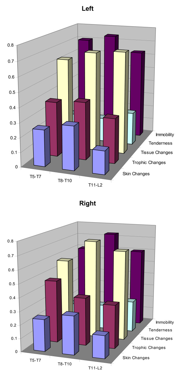
Methods: A case-control study was used to measure the association between type 2 diabetes mellitus and a series of 30 osteopathic palpatory findings. The latter included skin changes, trophic changes, tissue changes, tenderness, and immobility at spinal segmental levels T5-T7, T8-T10, and T11-L2 bilaterally. Logistic regression models that adjusted for age, sex, and comorbid conditions were used to compute odds ratios (ORs) and 95% confidence intervals (CIs) for the associations between type 2 diabetes mellitus and each of these findings.
Results and discussion: A total of 92 subjects were included in the study. After controlling for age, sex, hypertension, and clinical depression, the only significant finding was an association between type 2 diabetes mellitus and tissue changes at T11-L2 on the right side (OR, 5.54; 95% CI, 1.76-17.47; P = .003). Subgroup analyses of subjects with type 2 diabetes mellitus and hypertension demonstrated significant associations with tissue changes at T11-L2 bilaterally (OR, 27.38; 95% CI, 1.75-428; P = .02 for the left side and OR, 24.00; 95% CI, 1.51-382; P = .02 for the right side). Among subjects with type 2 diabetes mellitus and hypertension, there was also a strong diabetes mellitus duration effect for tissue changes at T11-L2 bilaterally (OR, 12.00; 95% CI, 1.02-141; P = .05 for short duration vs. OR, 32.00; 95% CI, 2.29-448; P = .01 for long duration on the left side; and OR, 17.33; 95% CI, 1.39-217; P = .03 for short duration vs. OR, 32.00; 95% CI, 2.29-448; P = .01 for long duration on the right side).
Conclusion: The only consistent finding in this study was an association between type 2 diabetes mellitus and tissue changes at T11-L2 on the right side. Potential explanations for this finding include reflex viscerosomatic changes directly related to the progression of type 2 diabetes mellitus, a spurious association attributable to confounding visceral diseases, or a chance observation unrelated to type 2 diabetes mellitus. Larger prospective studies are needed to better study osteopathic palpatory findings in type 2 diabetes mellitus.

 求助内容:
求助内容: 应助结果提醒方式:


