Zhuangyu Zhang, Josef Machac, Gerard Helft, Stephen G Worthley, Cheuk Tang, Azfar G Zaman, Oswaldo J Rodriguez, Monte S Buchsbaum, Valentin Fuster, Juan J Badimon
{"title":"Non-invasive imaging of atherosclerotic plaque macrophage in a rabbit model with F-18 FDG PET: a histopathological correlation.","authors":"Zhuangyu Zhang, Josef Machac, Gerard Helft, Stephen G Worthley, Cheuk Tang, Azfar G Zaman, Oswaldo J Rodriguez, Monte S Buchsbaum, Valentin Fuster, Juan J Badimon","doi":"10.1186/1471-2385-6-3","DOIUrl":null,"url":null,"abstract":"<p><strong>Background: </strong>Coronary atherosclerosis and its thrombotic complications are the major cause of mortality and morbidity throughout the industrialized world. Thrombosis on disrupted atherosclerotic plaques plays a key role in the onset of acute coronary syndromes. Macrophages density is one of the most critical compositions of plaque in both plaque vulnerability and thrombogenicity upon rupture. It has been shown that macrophages have a high uptake of 18F-FDG (FDG). We studied the correlation of FDG uptake with histopathological macrophage accumulation in atherosclerotic plaques in a rabbit model.</p><p><strong>Methods: </strong>Atherosclerosis was induced in rabbits (n = 6) by a combination of atherogenic diet and balloon denudation of the aorta. PET imaging was performed at baseline and 2 months after atherogenic diet and coregistered with magnetic resonance (MR) imaging. Normal (n = 3) rabbits served as controls. FDG uptake by the thoracic aorta was expressed as concentration (muCi/ml) and the ratio of aortic uptake-to-blood radioactivity. FDG uptake and RAM-11 antibody positive areas were analyzed in descending aorta.</p><p><strong>Results: </strong>Atherosclerotic aortas showed significantly higher uptake of FDG than normal aortas. The correlation of aortic FDG uptake with macrophage areas assessed by histopathology was statistically significant although it was not high (r = 0.48, p < 0.0001). When uptake was expressed as the ratio of aortic uptake-to-blood activity, it correlated better (r = 0.80, p < 0.0001) with the macrophage areas, due to the correction for residual blood FDG activity.</p><p><strong>Conclusion: </strong>PET FDG activity correlated with macrophage content within aortic atherosclerosis. This imaging approach might serve as a useful non-invasive imaging technique and potentially permit monitoring of relative changes in inflammation within the atherosclerotic lesion.</p>","PeriodicalId":80684,"journal":{"name":"BMC nuclear medicine","volume":"6 ","pages":"3"},"PeriodicalIF":0.0000,"publicationDate":"2006-05-25","publicationTypes":"Journal Article","fieldsOfStudy":null,"isOpenAccess":false,"openAccessPdf":"https://sci-hub-pdf.com/10.1186/1471-2385-6-3","citationCount":"126","resultStr":null,"platform":"Semanticscholar","paperid":null,"PeriodicalName":"BMC nuclear medicine","FirstCategoryId":"1085","ListUrlMain":"https://doi.org/10.1186/1471-2385-6-3","RegionNum":0,"RegionCategory":null,"ArticlePicture":[],"TitleCN":null,"AbstractTextCN":null,"PMCID":null,"EPubDate":"","PubModel":"","JCR":"","JCRName":"","Score":null,"Total":0}
引用次数: 126
Abstract
Background: Coronary atherosclerosis and its thrombotic complications are the major cause of mortality and morbidity throughout the industrialized world. Thrombosis on disrupted atherosclerotic plaques plays a key role in the onset of acute coronary syndromes. Macrophages density is one of the most critical compositions of plaque in both plaque vulnerability and thrombogenicity upon rupture. It has been shown that macrophages have a high uptake of 18F-FDG (FDG). We studied the correlation of FDG uptake with histopathological macrophage accumulation in atherosclerotic plaques in a rabbit model.
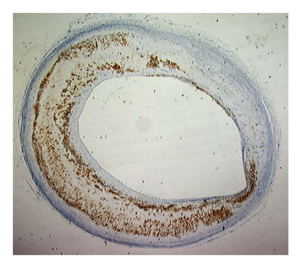
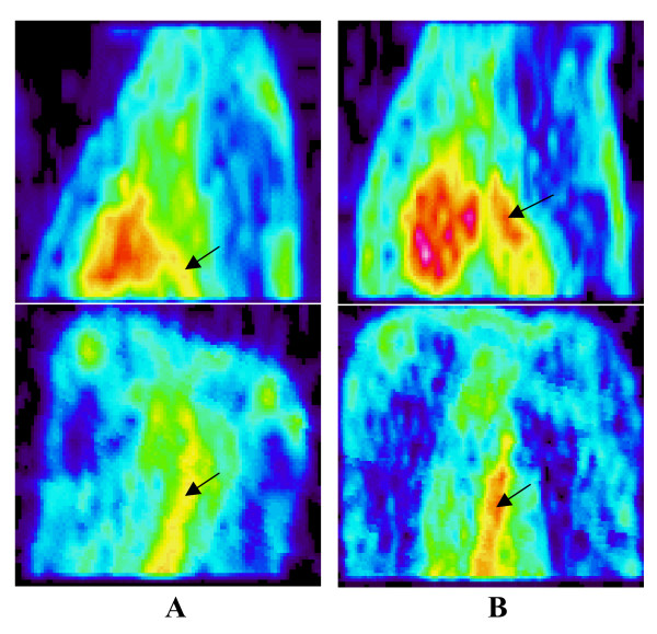
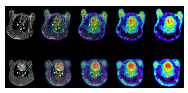
Methods: Atherosclerosis was induced in rabbits (n = 6) by a combination of atherogenic diet and balloon denudation of the aorta. PET imaging was performed at baseline and 2 months after atherogenic diet and coregistered with magnetic resonance (MR) imaging. Normal (n = 3) rabbits served as controls. FDG uptake by the thoracic aorta was expressed as concentration (muCi/ml) and the ratio of aortic uptake-to-blood radioactivity. FDG uptake and RAM-11 antibody positive areas were analyzed in descending aorta.
Results: Atherosclerotic aortas showed significantly higher uptake of FDG than normal aortas. The correlation of aortic FDG uptake with macrophage areas assessed by histopathology was statistically significant although it was not high (r = 0.48, p < 0.0001). When uptake was expressed as the ratio of aortic uptake-to-blood activity, it correlated better (r = 0.80, p < 0.0001) with the macrophage areas, due to the correction for residual blood FDG activity.
Conclusion: PET FDG activity correlated with macrophage content within aortic atherosclerosis. This imaging approach might serve as a useful non-invasive imaging technique and potentially permit monitoring of relative changes in inflammation within the atherosclerotic lesion.

 求助内容:
求助内容: 应助结果提醒方式:


