{"title":"Cytokine profiles of filarial granulomas in jirds infected with Brugia pahangi.","authors":"Ramakrishna U Rao, Thomas R Klei","doi":"10.1186/1475-2883-5-3","DOIUrl":null,"url":null,"abstract":"<p><strong>Background: </strong>A granulomatous inflammatory response develops in jirds infected subcutaneously or intraperitoneally with filarial nematodes namely Brugia pahangi and B. malayi. Previous studies by light and electron microscopy have shown cellular inflammatory responses in and around these granulomas. Furthermore, the cellular inflammatory responses of granulomas found in the lymphatics and peritoneal cavity appear to be similar. The purpose of this study was to determine the cytokine profiles of granulomas in the peritoneal cavity of B. pahangi-infected jirds and to determine whether the granulomas release any proinflammatory cytokines ex vivo.</p><p><strong>Methods: </strong>A semiautomated quantitative polymerase chain reaction (Q-PCR) was performed on cDNA prepared from the granulomas of infected jirds to study the species-specific mRNA expression of IL-2, IL-4, IFN-gamma, IL-5, and IL-10. Genomic DNA was extracted from the granulomas, and parasite DNA was detected by Q-PCR by amplifying the HhaI repeat sequence. The levels of the inflammation-causing cytokines IL-6 and TNFalpha that were secreted by the granulomas were measured by cell-based assays.</p><p><strong>Results: </strong>Florid granulomas showed higher levels of IFN-gamma than other cytokines, linking this Th1 cytokine to the granulomatous inflammation that develops in jirds and humans. IL-4 expression was much lower than that of IFN-gamma but higher than that of IL-10. A low level of IL-5 mRNA expression was detectable in all granulomas as was the level of IL-2 expression. The levels of the inflammatory cytokines IL-6 and TNFalpha, secreted by intact granulomas, spontaneously increased by 48 h after culture. Parasite antigen stimulation and subsequent release of IL-6 and TNFalpha by the granulomas indicated a moderate increase in the levels of these two cytokines. The amplification of the Brugia HhaI repeat DNA and Wolbachia 16S rDNA indicated worm components and bacterial components in the granulomatous tissue.</p><p><strong>Conclusion: </strong>Granuloma development in filarial infections is a complex process involving cellular reactions responding to parasite/bacteria and their components. The interactions between worm-derived granulomas and their hosts are dynamic and multifaceted. The data collected thus far suggest that the expression profiles of many of the measured cytokines in the lymphoid tissues of Brugia-infected jirds are different from those of the cytokines in granulomas. Moreover, granulomas have the ability to secrete the inflammatory cytokines IL-6 and TNFalpha.</p>","PeriodicalId":84756,"journal":{"name":"Filaria journal","volume":"5 ","pages":"3"},"PeriodicalIF":0.0000,"publicationDate":"2006-03-16","publicationTypes":"Journal Article","fieldsOfStudy":null,"isOpenAccess":false,"openAccessPdf":"https://sci-hub-pdf.com/10.1186/1475-2883-5-3","citationCount":"9","resultStr":null,"platform":"Semanticscholar","paperid":null,"PeriodicalName":"Filaria journal","FirstCategoryId":"1085","ListUrlMain":"https://doi.org/10.1186/1475-2883-5-3","RegionNum":0,"RegionCategory":null,"ArticlePicture":[],"TitleCN":null,"AbstractTextCN":null,"PMCID":null,"EPubDate":"","PubModel":"","JCR":"","JCRName":"","Score":null,"Total":0}
引用次数: 9
Abstract
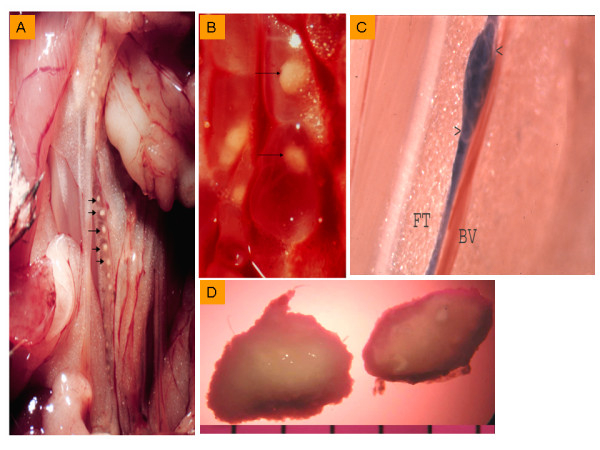
Background: A granulomatous inflammatory response develops in jirds infected subcutaneously or intraperitoneally with filarial nematodes namely Brugia pahangi and B. malayi. Previous studies by light and electron microscopy have shown cellular inflammatory responses in and around these granulomas. Furthermore, the cellular inflammatory responses of granulomas found in the lymphatics and peritoneal cavity appear to be similar. The purpose of this study was to determine the cytokine profiles of granulomas in the peritoneal cavity of B. pahangi-infected jirds and to determine whether the granulomas release any proinflammatory cytokines ex vivo.
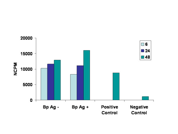
Methods: A semiautomated quantitative polymerase chain reaction (Q-PCR) was performed on cDNA prepared from the granulomas of infected jirds to study the species-specific mRNA expression of IL-2, IL-4, IFN-gamma, IL-5, and IL-10. Genomic DNA was extracted from the granulomas, and parasite DNA was detected by Q-PCR by amplifying the HhaI repeat sequence. The levels of the inflammation-causing cytokines IL-6 and TNFalpha that were secreted by the granulomas were measured by cell-based assays.
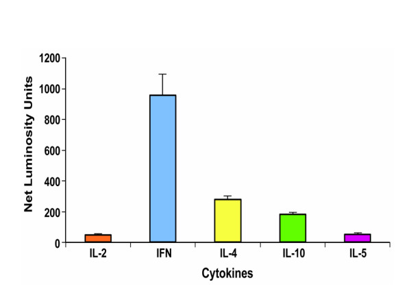
Results: Florid granulomas showed higher levels of IFN-gamma than other cytokines, linking this Th1 cytokine to the granulomatous inflammation that develops in jirds and humans. IL-4 expression was much lower than that of IFN-gamma but higher than that of IL-10. A low level of IL-5 mRNA expression was detectable in all granulomas as was the level of IL-2 expression. The levels of the inflammatory cytokines IL-6 and TNFalpha, secreted by intact granulomas, spontaneously increased by 48 h after culture. Parasite antigen stimulation and subsequent release of IL-6 and TNFalpha by the granulomas indicated a moderate increase in the levels of these two cytokines. The amplification of the Brugia HhaI repeat DNA and Wolbachia 16S rDNA indicated worm components and bacterial components in the granulomatous tissue.
Conclusion: Granuloma development in filarial infections is a complex process involving cellular reactions responding to parasite/bacteria and their components. The interactions between worm-derived granulomas and their hosts are dynamic and multifaceted. The data collected thus far suggest that the expression profiles of many of the measured cytokines in the lymphoid tissues of Brugia-infected jirds are different from those of the cytokines in granulomas. Moreover, granulomas have the ability to secrete the inflammatory cytokines IL-6 and TNFalpha.

 求助内容:
求助内容: 应助结果提醒方式:


