Mohammad Eftekhari, Majid Assadi, Majid Kazemi, Mohsen Saghari, Armaghan Fard Esfahani, Babak Fallahi Sichani, Ali Gholamrezanezhad, Davood Beiki
{"title":"A preliminary study of neuroSPECT evaluation of patients with post-traumatic smell impairment.","authors":"Mohammad Eftekhari, Majid Assadi, Majid Kazemi, Mohsen Saghari, Armaghan Fard Esfahani, Babak Fallahi Sichani, Ali Gholamrezanezhad, Davood Beiki","doi":"10.1186/1471-2385-5-6","DOIUrl":null,"url":null,"abstract":"<p><strong>Background: </strong>Most olfactory testings are subjective and since they depend upon the patients' response, they are prone to false positive results. The aim of this study was to use quantitative brain perfusion SPECT in order to detect possible areas of brain activation in response to odorant stimulation in patients with post-traumatic impaired smell in comparison to a group of normal subjects.</p><p><strong>Methods: </strong>Fourteen patients with post-traumatic impaired smell and ten healthy controls were entered in this prospective study. All subjects underwent brain SPECT after intravenous injection of 740-MBq 99mTc-ECD and 48 hours later, the same procedure was repeated following olfactory stimulus (vanilla powder).</p><p><strong>Results: </strong>In most of seven regions of interest (Orbital Frontal Cortex, Inferior Frontal Pole, Superior Frontal Pole, Posterior Superior Frontal Lobe, Parasagittal Area, Occipital Pole, and Cerebellar area) the post-stimulation quantitative values show increased cortical perfusion being more pronounced in normal volunteers than the anosmic patients (except cerebellar areas and the right occipital pole). Maximal activation was observed in orbitofrontal regions (right+ 25.45% and left +25.47%).</p><p><strong>Conclusion: </strong>Brain SPECT is a valuable imaging technique in the assessment of post-traumatic anosmia and could be competitive as an alternative to other imaging techniques, especially when functional MRI is unavailable or unsuitable. However, this procedure may benefit from complementary MRI or CT anatomical imaging.</p>","PeriodicalId":80684,"journal":{"name":"BMC nuclear medicine","volume":"5 ","pages":"6"},"PeriodicalIF":0.0000,"publicationDate":"2005-11-28","publicationTypes":"Journal Article","fieldsOfStudy":null,"isOpenAccess":false,"openAccessPdf":"https://sci-hub-pdf.com/10.1186/1471-2385-5-6","citationCount":"15","resultStr":null,"platform":"Semanticscholar","paperid":null,"PeriodicalName":"BMC nuclear medicine","FirstCategoryId":"1085","ListUrlMain":"https://doi.org/10.1186/1471-2385-5-6","RegionNum":0,"RegionCategory":null,"ArticlePicture":[],"TitleCN":null,"AbstractTextCN":null,"PMCID":null,"EPubDate":"","PubModel":"","JCR":"","JCRName":"","Score":null,"Total":0}
引用次数: 15
Abstract
Background: Most olfactory testings are subjective and since they depend upon the patients' response, they are prone to false positive results. The aim of this study was to use quantitative brain perfusion SPECT in order to detect possible areas of brain activation in response to odorant stimulation in patients with post-traumatic impaired smell in comparison to a group of normal subjects.
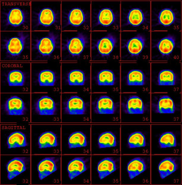
Methods: Fourteen patients with post-traumatic impaired smell and ten healthy controls were entered in this prospective study. All subjects underwent brain SPECT after intravenous injection of 740-MBq 99mTc-ECD and 48 hours later, the same procedure was repeated following olfactory stimulus (vanilla powder).
Results: In most of seven regions of interest (Orbital Frontal Cortex, Inferior Frontal Pole, Superior Frontal Pole, Posterior Superior Frontal Lobe, Parasagittal Area, Occipital Pole, and Cerebellar area) the post-stimulation quantitative values show increased cortical perfusion being more pronounced in normal volunteers than the anosmic patients (except cerebellar areas and the right occipital pole). Maximal activation was observed in orbitofrontal regions (right+ 25.45% and left +25.47%).
Conclusion: Brain SPECT is a valuable imaging technique in the assessment of post-traumatic anosmia and could be competitive as an alternative to other imaging techniques, especially when functional MRI is unavailable or unsuitable. However, this procedure may benefit from complementary MRI or CT anatomical imaging.


 求助内容:
求助内容: 应助结果提醒方式:


