Il-Jung Park, Youn-Tae Roh, Seung-Han Shin, Ho-Yeon Park, Changhoon Jeong, Soo-Hwan Kang
{"title":"Importance of detection of capitellar cartilage injuries concomitant with isolated radial head fractures: A retrospective clinical study.","authors":"Il-Jung Park, Youn-Tae Roh, Seung-Han Shin, Ho-Yeon Park, Changhoon Jeong, Soo-Hwan Kang","doi":"10.5152/j.aott.2021.20046","DOIUrl":null,"url":null,"abstract":"<p><strong>Objective: </strong>This study aimed to analyze the injury pattern and clinical importance of concomitant capitellar cartilage defects (CCDs) among patients treated surgically for radial head fracture (RHF).</p><p><strong>Methods: </strong>A total of 74 patients who were treated surgically for isolated RHFs were retrospectively reviewed. Of these, 12 patients with CCDs (16.2%) were classified as Group I (10 men; mean age, 41.3±12.8 years) and the remaining 62 patients without CCD as Group II (control group) (48 men; mean age, 50.8±13 years). The mean follow-up was 21.3±3.2 months in Group I and 18.7±6.4 in Group II. In Group I, 11 patients underwent open reduction and internal fixation, whereas 1 patient was treated by radial head resection. The preoperative range of motion (ROM) was recorded; the severity of RHF was assessed using the Mason classification. The location, size, and thickness of CCD injuries at the time of surgery were also documented. At the final follow-up, radiological assessment was performed to determine the bone union, and clinical measurements, including ROM and the Mayo elbow performance score (MEPS), were performed. The clinical features of the 2 groups were statistically analyzed.</p><p><strong>Results: </strong>In Group I, 10 patients showed limited forearm rotation. CCD was located posterolaterally in 11 patients and anterolaterally in 1 patient. At the final follow-up, 11 patients from Group I who underwent open reduction and internal fixation showed complete union of RHF and full recovery of pronation and supination. According to the MEPS, 9 patients exhibited excellent results, and 3 patients exhibited good results. In Group I, RHFs were classified as Mason type II in 7 patients (58.3%) and type III in 4 patients (58.3%). In Group II, RHFs were type II in 45 patients (72.6%) and type III in 17 patients (27.4%). In comparative analyses, there was a significant difference in age (41.3±12.8 versus 50.8±13.0, p=0.041) between the 2 groups. Preoperative pronation/supination was higher in Group II (131.7±36.2) than in Group I (106.3±31.6) (p=0.021). There were no significant differences in sex (p=0.097), follow-up period (p=0.326), Mason type (p=0.482), preoperative extension/flexion (102.3±43.3 [Group I] versus 107.6±44.9 [Group II]) (p=0.584), final follow-up extension/flexion (133.3±10.7 [Group I] versus 126.9±21.2 [Group II]) (p=0.384), pronation/supination (151.2±9.1 [Group I] versus 151.2±13.3 [Group II]) (p=0.558), and the MEPSs (92.9±6.6 [Group I] versus 93.3±7.5 [Group II]) (p=0.701).</p><p><strong>Conclusion: </strong>If a thorough physical examination of a patient with RHF reveals limited forearm rotation, effort must be made to identify the cause, and the possibility of CCD must be considered. Moreover, there is a need for careful observation during RHF surgery for not only fracture reduction or fixation but also possible CCD.</p><p><strong>Level of evidence: </strong>Level III, Therapeutic Study.</p>","PeriodicalId":7097,"journal":{"name":"Acta orthopaedica et traumatologica turcica","volume":"55 2","pages":"112-117"},"PeriodicalIF":1.1000,"publicationDate":"2021-03-01","publicationTypes":"Journal Article","fieldsOfStudy":null,"isOpenAccess":false,"openAccessPdf":"https://www.ncbi.nlm.nih.gov/pmc/articles/PMC11229612/pdf/","citationCount":"0","resultStr":null,"platform":"Semanticscholar","paperid":null,"PeriodicalName":"Acta orthopaedica et traumatologica turcica","FirstCategoryId":"3","ListUrlMain":"https://doi.org/10.5152/j.aott.2021.20046","RegionNum":4,"RegionCategory":"医学","ArticlePicture":[],"TitleCN":null,"AbstractTextCN":null,"PMCID":null,"EPubDate":"","PubModel":"","JCR":"Q3","JCRName":"ORTHOPEDICS","Score":null,"Total":0}
引用次数: 0
Abstract
Objective: This study aimed to analyze the injury pattern and clinical importance of concomitant capitellar cartilage defects (CCDs) among patients treated surgically for radial head fracture (RHF).
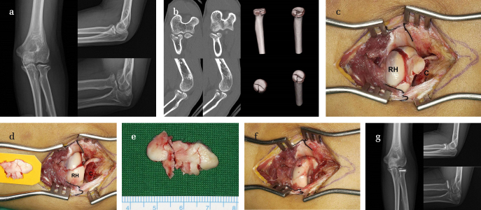
Methods: A total of 74 patients who were treated surgically for isolated RHFs were retrospectively reviewed. Of these, 12 patients with CCDs (16.2%) were classified as Group I (10 men; mean age, 41.3±12.8 years) and the remaining 62 patients without CCD as Group II (control group) (48 men; mean age, 50.8±13 years). The mean follow-up was 21.3±3.2 months in Group I and 18.7±6.4 in Group II. In Group I, 11 patients underwent open reduction and internal fixation, whereas 1 patient was treated by radial head resection. The preoperative range of motion (ROM) was recorded; the severity of RHF was assessed using the Mason classification. The location, size, and thickness of CCD injuries at the time of surgery were also documented. At the final follow-up, radiological assessment was performed to determine the bone union, and clinical measurements, including ROM and the Mayo elbow performance score (MEPS), were performed. The clinical features of the 2 groups were statistically analyzed.
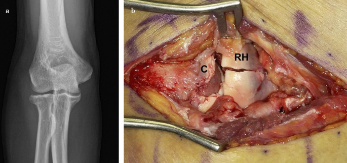
Results: In Group I, 10 patients showed limited forearm rotation. CCD was located posterolaterally in 11 patients and anterolaterally in 1 patient. At the final follow-up, 11 patients from Group I who underwent open reduction and internal fixation showed complete union of RHF and full recovery of pronation and supination. According to the MEPS, 9 patients exhibited excellent results, and 3 patients exhibited good results. In Group I, RHFs were classified as Mason type II in 7 patients (58.3%) and type III in 4 patients (58.3%). In Group II, RHFs were type II in 45 patients (72.6%) and type III in 17 patients (27.4%). In comparative analyses, there was a significant difference in age (41.3±12.8 versus 50.8±13.0, p=0.041) between the 2 groups. Preoperative pronation/supination was higher in Group II (131.7±36.2) than in Group I (106.3±31.6) (p=0.021). There were no significant differences in sex (p=0.097), follow-up period (p=0.326), Mason type (p=0.482), preoperative extension/flexion (102.3±43.3 [Group I] versus 107.6±44.9 [Group II]) (p=0.584), final follow-up extension/flexion (133.3±10.7 [Group I] versus 126.9±21.2 [Group II]) (p=0.384), pronation/supination (151.2±9.1 [Group I] versus 151.2±13.3 [Group II]) (p=0.558), and the MEPSs (92.9±6.6 [Group I] versus 93.3±7.5 [Group II]) (p=0.701).
Conclusion: If a thorough physical examination of a patient with RHF reveals limited forearm rotation, effort must be made to identify the cause, and the possibility of CCD must be considered. Moreover, there is a need for careful observation during RHF surgery for not only fracture reduction or fixation but also possible CCD.
期刊介绍:
Acta Orthopaedica et Traumatologica Turcica (AOTT) is an international, scientific, open access periodical published in accordance with independent, unbiased, and double-blinded peer-review principles. The journal is the official publication of the Turkish Association of Orthopaedics and Traumatology, and Turkish Society of Orthopaedics and Traumatology. It is published bimonthly in January, March, May, July, September, and November. The publication language of the journal is English.
The aim of the journal is to publish original studies of the highest scientific and clinical value in orthopedics, traumatology, and related disciplines. The scope of the journal includes but not limited to diagnostic, treatment, and prevention methods related to orthopedics and traumatology. Acta Orthopaedica et Traumatologica Turcica publishes clinical and basic research articles, case reports, personal clinical and technical notes, systematic reviews and meta-analyses and letters to the Editor. Proceedings of scientific meetings are also considered for publication.
The target audience of the journal includes healthcare professionals, physicians, and researchers who are interested or working in orthopedics and traumatology field, and related disciplines.
 求助内容:
求助内容: 应助结果提醒方式:


