{"title":"Ventriculocoronary Fistulas with Hypoplastic Left Heart in a Neonate: Imaging with Cardiac CT.","authors":"Serap Baş, Utku Alkara","doi":"10.1155/2021/6657447","DOIUrl":null,"url":null,"abstract":"<p><p>Fistulous communications between the ventricular cavities and the coronary arterial tree can be found in the presence of hypoplasia of the left ventricle, especially when the ventricular septum is intact and mitral stenosis and aortic atresia subtype are present. The cardiac CT provides excellent anatomic information especially in the evaluation of extracardiac vessels and coronary arteries. In this case study, we report a newborn with ventriculocoronary fistulas (VCFs) with the hypoplastic left disease diagnosed with cardiac CT. Transthoracic echocardiography of a term baby showed hypoplastic left heart syndrome (HLHS) with mitral stenosis and aortic atresia. The patient immediately underwent a Sano variation of the Norwood procedure. On the postoperative second day, the clinical status of the patient deteriorated. A prospective electrocardiogram-gated axial technique was performed within a single heartbeat for the patient and large VCFs were detected and a second operation were performed to close the VCFs that failed. On the nineteenth day after the operation, the baby passed away. According to us, cardiac CT can also be performed free-breathing and without anesthesia in the neonatal period for the definition of complex cardiac anatomy with the lower radiation dose from the latest scanners, radiation risk of CT should be weighed against the anesthesia risk of cardiac MRI and intraoperative risk of conventional cardiac angiography. Pre-operative cardiac CT may increase surgical success.</p>","PeriodicalId":30326,"journal":{"name":"Case Reports in Radiology","volume":" ","pages":"6657447"},"PeriodicalIF":0.0000,"publicationDate":"2021-03-16","publicationTypes":"Journal Article","fieldsOfStudy":null,"isOpenAccess":false,"openAccessPdf":"https://www.ncbi.nlm.nih.gov/pmc/articles/PMC7987453/pdf/","citationCount":"0","resultStr":null,"platform":"Semanticscholar","paperid":null,"PeriodicalName":"Case Reports in Radiology","FirstCategoryId":"1085","ListUrlMain":"https://doi.org/10.1155/2021/6657447","RegionNum":0,"RegionCategory":null,"ArticlePicture":[],"TitleCN":null,"AbstractTextCN":null,"PMCID":null,"EPubDate":"2021/1/1 0:00:00","PubModel":"eCollection","JCR":"","JCRName":"","Score":null,"Total":0}
引用次数: 0
Abstract
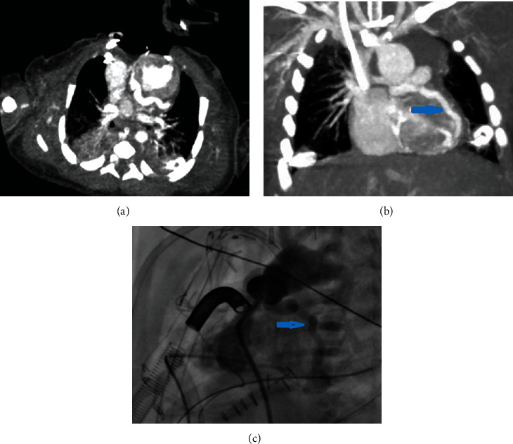
Fistulous communications between the ventricular cavities and the coronary arterial tree can be found in the presence of hypoplasia of the left ventricle, especially when the ventricular septum is intact and mitral stenosis and aortic atresia subtype are present. The cardiac CT provides excellent anatomic information especially in the evaluation of extracardiac vessels and coronary arteries. In this case study, we report a newborn with ventriculocoronary fistulas (VCFs) with the hypoplastic left disease diagnosed with cardiac CT. Transthoracic echocardiography of a term baby showed hypoplastic left heart syndrome (HLHS) with mitral stenosis and aortic atresia. The patient immediately underwent a Sano variation of the Norwood procedure. On the postoperative second day, the clinical status of the patient deteriorated. A prospective electrocardiogram-gated axial technique was performed within a single heartbeat for the patient and large VCFs were detected and a second operation were performed to close the VCFs that failed. On the nineteenth day after the operation, the baby passed away. According to us, cardiac CT can also be performed free-breathing and without anesthesia in the neonatal period for the definition of complex cardiac anatomy with the lower radiation dose from the latest scanners, radiation risk of CT should be weighed against the anesthesia risk of cardiac MRI and intraoperative risk of conventional cardiac angiography. Pre-operative cardiac CT may increase surgical success.

 求助内容:
求助内容: 应助结果提醒方式:


