Carmen Sandoval, Gabriela Araujo, Wilfredo Sosa, Sara Avalos, Fernando Silveira, Carlos Corbett, Concepción Zúniga, Marcia Laurenti
{"title":"<i>In situ</i> cellular immune response in non-ulcerated skin lesions due to <i>Leishmania (L.) infantum chagasi</i> infection.","authors":"Carmen Sandoval, Gabriela Araujo, Wilfredo Sosa, Sara Avalos, Fernando Silveira, Carlos Corbett, Concepción Zúniga, Marcia Laurenti","doi":"10.1590/1678-9199-JVATITD-2020-0149","DOIUrl":null,"url":null,"abstract":"<p><strong>Background: </strong>Skin lesions of patients affected by non-ulcerated cutaneous leishmaniasis (NUCL) caused by <i>L. (L.) infantum chagasi</i> are characterized by lymphohistiocytic inflammatory infiltrate associated with epithelioid granuloma and scarce parasitism. However, the <i>in situ</i> cellular immune response of these patients is unclear. Therefore, the aim of the present study was to characterize the cellular immune response in the skin lesions of patients affected by NUCL.</p><p><strong>Methods: </strong>Twenty biopsies were processed by immunohistochemistry using primary antibodies to T lymphocytes (CD4, CD8), NK cells, B lymphocytes, macrophages, nitric oxide synthase and interferon-gamma.</p><p><strong>Results: </strong>Immunohistochemistry revealed higher expression of all cellular types and molecules (IFN-γ, iNOS) in the dermis of diseased skin compared to the skin of healthy individuals (p < 0.05). Morphometric analysis performed in the skin lesions sections showed the predominance of CD8<sup>+</sup> T lymphocytes in the mononuclear infiltrate, followed by macrophages, mostly iNOS<sup>+</sup>, a response that could be mediated by IFN-γ.</p><p><strong>Conclusion: </strong>Our study improves knowledge of the cellular immune response in non-ulcerated or atypical cutaneous leishmaniasis caused by <i>L. (L.) infantum chagasi</i> in Central America and pointed to the pivotal participation of CD8<sup>+</sup> T lymphocytes in the host defense mechanisms against the parasite in patients with NUCL.</p>","PeriodicalId":520810,"journal":{"name":"The journal of venomous animals and toxins including tropical diseases","volume":" ","pages":"e20200149"},"PeriodicalIF":1.8000,"publicationDate":"2021-02-26","publicationTypes":"Journal Article","fieldsOfStudy":null,"isOpenAccess":false,"openAccessPdf":"https://www.ncbi.nlm.nih.gov/pmc/articles/PMC7909480/pdf/","citationCount":"7","resultStr":null,"platform":"Semanticscholar","paperid":null,"PeriodicalName":"The journal of venomous animals and toxins including tropical diseases","FirstCategoryId":"3","ListUrlMain":"https://doi.org/10.1590/1678-9199-JVATITD-2020-0149","RegionNum":0,"RegionCategory":null,"ArticlePicture":[],"TitleCN":null,"AbstractTextCN":null,"PMCID":null,"EPubDate":"2021/1/1 0:00:00","PubModel":"eCollection","JCR":"","JCRName":"","Score":null,"Total":0}
引用次数: 7
Abstract
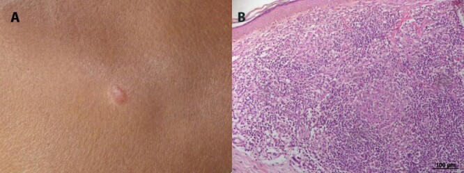
Background: Skin lesions of patients affected by non-ulcerated cutaneous leishmaniasis (NUCL) caused by L. (L.) infantum chagasi are characterized by lymphohistiocytic inflammatory infiltrate associated with epithelioid granuloma and scarce parasitism. However, the in situ cellular immune response of these patients is unclear. Therefore, the aim of the present study was to characterize the cellular immune response in the skin lesions of patients affected by NUCL.
Methods: Twenty biopsies were processed by immunohistochemistry using primary antibodies to T lymphocytes (CD4, CD8), NK cells, B lymphocytes, macrophages, nitric oxide synthase and interferon-gamma.
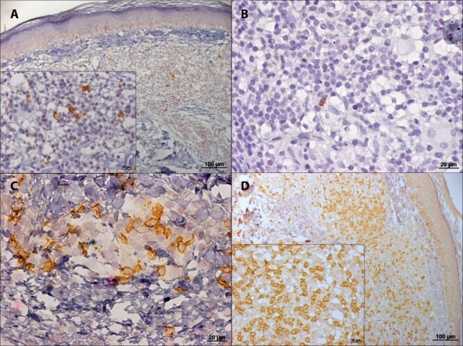
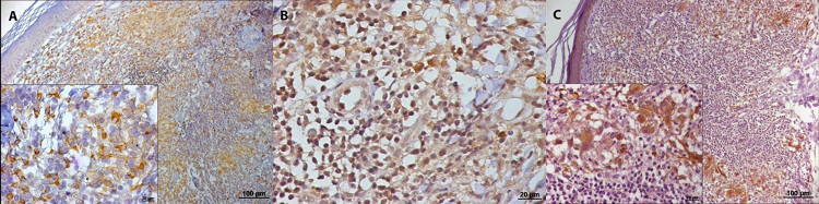
Results: Immunohistochemistry revealed higher expression of all cellular types and molecules (IFN-γ, iNOS) in the dermis of diseased skin compared to the skin of healthy individuals (p < 0.05). Morphometric analysis performed in the skin lesions sections showed the predominance of CD8+ T lymphocytes in the mononuclear infiltrate, followed by macrophages, mostly iNOS+, a response that could be mediated by IFN-γ.
Conclusion: Our study improves knowledge of the cellular immune response in non-ulcerated or atypical cutaneous leishmaniasis caused by L. (L.) infantum chagasi in Central America and pointed to the pivotal participation of CD8+ T lymphocytes in the host defense mechanisms against the parasite in patients with NUCL.

 求助内容:
求助内容: 应助结果提醒方式:


