Siroos Mirzaei, Michel Guerchaft, Christopher Bonnier, Peter Knoll, Michel Doat, Peter Braeutigam
{"title":"Use of segmented CT transmission map to avoid metal artifacts in PET images by a PET-CT device.","authors":"Siroos Mirzaei, Michel Guerchaft, Christopher Bonnier, Peter Knoll, Michel Doat, Peter Braeutigam","doi":"10.1186/1471-2385-5-3","DOIUrl":null,"url":null,"abstract":"<p><p>BACKGROUND: Attenuation correction is generally used to PET images to achieve count rate values independent from tissue densities. The goal of this study was to provide a qualitative comparison of attenuation corrected PET images produced by a PET-CT device (CT, 120 kV, 40 mAs, FOV 600 mm) with and without segmentation of transmission data (ACseg+ and ACseg-respectively). Methods: The reconstructed images were compared to attenuation corrected images obtained with a high-energy transmission source (Cs-137 - 662 keV).Thirty oncologic patients were studied using CT and 137Cs for attenuation correction. All image data were acquired using the Gemini PET-CT scanner (Philips Medical Systems). It is an open PET-CT system that consists of the MX8000 multislice CT and the Allegro PET scanner arranged in a separable configuration. Images with ACseg+ and ACseg- were analyzed simultaneously in coronal, sagittal and transaxial planes. Two nuclear medicine physicians reviewed the image sets. Results: The image quality in the area of metal implants was better with ACseg+ than ACseg-, without metal induced artifacts generally observed in CT corrected images. Further the images with ACseg+ were qualitatively comparable to those obtained with 137Cs attenuation correction. Conclusions: In case of metal implants, PET studies corrected by CT should preferably use the ACseg+ method to avoid the image artifacts.</p>","PeriodicalId":80684,"journal":{"name":"BMC nuclear medicine","volume":"5 1","pages":"3"},"PeriodicalIF":0.0000,"publicationDate":"2005-06-14","publicationTypes":"Journal Article","fieldsOfStudy":null,"isOpenAccess":false,"openAccessPdf":"https://sci-hub-pdf.com/10.1186/1471-2385-5-3","citationCount":"38","resultStr":null,"platform":"Semanticscholar","paperid":null,"PeriodicalName":"BMC nuclear medicine","FirstCategoryId":"1085","ListUrlMain":"https://doi.org/10.1186/1471-2385-5-3","RegionNum":0,"RegionCategory":null,"ArticlePicture":[],"TitleCN":null,"AbstractTextCN":null,"PMCID":null,"EPubDate":"","PubModel":"","JCR":"","JCRName":"","Score":null,"Total":0}
引用次数: 38
Abstract
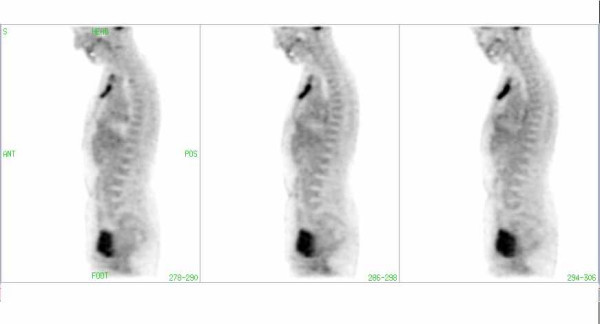
BACKGROUND: Attenuation correction is generally used to PET images to achieve count rate values independent from tissue densities. The goal of this study was to provide a qualitative comparison of attenuation corrected PET images produced by a PET-CT device (CT, 120 kV, 40 mAs, FOV 600 mm) with and without segmentation of transmission data (ACseg+ and ACseg-respectively). Methods: The reconstructed images were compared to attenuation corrected images obtained with a high-energy transmission source (Cs-137 - 662 keV).Thirty oncologic patients were studied using CT and 137Cs for attenuation correction. All image data were acquired using the Gemini PET-CT scanner (Philips Medical Systems). It is an open PET-CT system that consists of the MX8000 multislice CT and the Allegro PET scanner arranged in a separable configuration. Images with ACseg+ and ACseg- were analyzed simultaneously in coronal, sagittal and transaxial planes. Two nuclear medicine physicians reviewed the image sets. Results: The image quality in the area of metal implants was better with ACseg+ than ACseg-, without metal induced artifacts generally observed in CT corrected images. Further the images with ACseg+ were qualitatively comparable to those obtained with 137Cs attenuation correction. Conclusions: In case of metal implants, PET studies corrected by CT should preferably use the ACseg+ method to avoid the image artifacts.


 求助内容:
求助内容: 应助结果提醒方式:


