Diabetes Severity in Cornea Donors and 1-Year Outcomes in Recipients in the Diabetes Endothelial Keratoplasty Study: A Secondary Analysis of a Randomized Clinical Trial.
Mark A Greiner, Vincent M Monnier, Baha M Arafah, Loretta B Szczotka-Flynn, Beth Ann Benetz, Ryan J Bailey, Robin L Gal, Craig Kollman, Keith H Baratz, James M Huffman, Ula V Jurkunas, Christopher S Ketcherside, Francis W Price, Gregory A Schmidt, Evan D Schoenberg, Mark C Soper, Lawrence R Tenkman, David D Verdier, Deepitha Nelson, David R Sell, Roy W Beck, Marianne O Price, Jonathan H Lass
{"title":"Diabetes Severity in Cornea Donors and 1-Year Outcomes in Recipients in the Diabetes Endothelial Keratoplasty Study: A Secondary Analysis of a Randomized Clinical Trial.","authors":"Mark A Greiner, Vincent M Monnier, Baha M Arafah, Loretta B Szczotka-Flynn, Beth Ann Benetz, Ryan J Bailey, Robin L Gal, Craig Kollman, Keith H Baratz, James M Huffman, Ula V Jurkunas, Christopher S Ketcherside, Francis W Price, Gregory A Schmidt, Evan D Schoenberg, Mark C Soper, Lawrence R Tenkman, David D Verdier, Deepitha Nelson, David R Sell, Roy W Beck, Marianne O Price, Jonathan H Lass","doi":"10.1001/jamaophthalmol.2026.4253","DOIUrl":null,"url":null,"abstract":"<p><strong>Importance: </strong>The effect of cornea donor diabetes severity on graft success, endothelial cell density (ECD), and morphometry after Descemet membrane endothelial keratoplasty (DMEK) is not known.</p><p><strong>Objective: </strong>To determine whether graft success, corneal endothelial cell loss (ECL), and morphometric changes 1 year after DMEK are associated with cornea donor diabetes severity measured by severity score, donor's postmortem level of hemoglobin A1c (HbA1c), and postmortem advanced glycation end products (AGEs) or advanced oxidation end products (AOPs).</p><p><strong>Design, setting, and participants: </strong>This was a secondary analysis of a multicenter, double-masked, randomized clinical trial at 28 clinical sites (46 surgeons) and 13 eye banks in the US among individuals undergoing low- to moderate-risk DMEK. AGE and AOP measurements were collected from a subset of donors. Data analysis was conducted from September 1, 2025, through April 30, 2026.</p><p><strong>Intervention: </strong>DMEK performed with a cornea from a donor without or with diabetes, assigned using a minimization procedure (comparable to randomization) to achieve an approximate 2:1 distribution, respectively.</p><p><strong>Main outcomes and measures: </strong>Graft success, ECL, coefficient of variation (CV) of cell area, and percentage of hexagonal cells (HEX) at 1 year from eye bank and postoperative specular central endothelial images.</p><p><strong>Results: </strong>A total of 1097 individuals (1421 study eyes; median [IQR] age, 71 [66-76] years; 631 [57.5%] female) undergoing low- to moderate-risk DMEK (1044 [95.2%] for Fuchs endothelial corneal dystrophy) were included. AGE and AOP measurements were collected from a subset of 472 donors (542 study donor corneas). Among 968 eyes with a postmortem donor HbA1c level lower than 6.5%, the 1-year graft success rate was 97.1% (95% CI, 95.4%-98.4%), compared with 97.4% (95% CI, 93.9%-100.0%), 93.0% (95% CI, 83.3%-98.8%), and 95.8% (95% CI, 87.3%-100.0%) among eyes with a donor HbA1c level of 6.5% to less than 7.5% (n = 194), 7.5% to less than 9.0% (n = 86), and 9.0% or higher (n = 48), respectively (P = .72). There was no significant difference in mean ECD across donor HbA1c groups. Donor diabetes severity measured with severity score, donor HbA1c level, and AGE and AOP metrics was not associated with 1-year graft failure, ECD, CV in cell area, or HEX percentage.</p><p><strong>Conclusions and relevance: </strong>This secondary analysis of a randomized clinical trial found that ECL and morphometry 1 year after DMEK were not affected by cornea donor diabetes severity. Comparable 1-year graft success, ECL, and cell morphometrics with tissue from donors with and without diabetes, regardless of the diabetes severity biomarker values observed, support the use of corneas from donors with diabetes for endothelial keratoplasty procedures without additional preoperative testing or risk stratification based on the diabetes status of the cornea donor.</p><p><strong>Trial registration: </strong>ClinicalTrials.gov Identifier: NCT05134480.</p>","PeriodicalId":14518,"journal":{"name":"JAMA ophthalmology","volume":" ","pages":""},"PeriodicalIF":10.5000,"publicationDate":"2026-10-08","publicationTypes":"Journal Article","fieldsOfStudy":null,"isOpenAccess":false,"openAccessPdf":"https://www.ncbi.nlm.nih.gov/pmc/articles/PMC13650705/pdf/","citationCount":"0","resultStr":null,"platform":"Semanticscholar","paperid":null,"PeriodicalName":"JAMA ophthalmology","FirstCategoryId":"3","ListUrlMain":"https://doi.org/10.1001/jamaophthalmol.2026.4253","RegionNum":1,"RegionCategory":"医学","ArticlePicture":[],"TitleCN":null,"AbstractTextCN":null,"PMCID":null,"EPubDate":"","PubModel":"","JCR":"Q1","JCRName":"OPHTHALMOLOGY","Score":null,"Total":0}
引用次数: 0
Abstract
Importance: The effect of cornea donor diabetes severity on graft success, endothelial cell density (ECD), and morphometry after Descemet membrane endothelial keratoplasty (DMEK) is not known.
Objective: To determine whether graft success, corneal endothelial cell loss (ECL), and morphometric changes 1 year after DMEK are associated with cornea donor diabetes severity measured by severity score, donor's postmortem level of hemoglobin A1c (HbA1c), and postmortem advanced glycation end products (AGEs) or advanced oxidation end products (AOPs).
Design, setting, and participants: This was a secondary analysis of a multicenter, double-masked, randomized clinical trial at 28 clinical sites (46 surgeons) and 13 eye banks in the US among individuals undergoing low- to moderate-risk DMEK. AGE and AOP measurements were collected from a subset of donors. Data analysis was conducted from September 1, 2025, through April 30, 2026.
Intervention: DMEK performed with a cornea from a donor without or with diabetes, assigned using a minimization procedure (comparable to randomization) to achieve an approximate 2:1 distribution, respectively.
Main outcomes and measures: Graft success, ECL, coefficient of variation (CV) of cell area, and percentage of hexagonal cells (HEX) at 1 year from eye bank and postoperative specular central endothelial images.
Results: A total of 1097 individuals (1421 study eyes; median [IQR] age, 71 [66-76] years; 631 [57.5%] female) undergoing low- to moderate-risk DMEK (1044 [95.2%] for Fuchs endothelial corneal dystrophy) were included. AGE and AOP measurements were collected from a subset of 472 donors (542 study donor corneas). Among 968 eyes with a postmortem donor HbA1c level lower than 6.5%, the 1-year graft success rate was 97.1% (95% CI, 95.4%-98.4%), compared with 97.4% (95% CI, 93.9%-100.0%), 93.0% (95% CI, 83.3%-98.8%), and 95.8% (95% CI, 87.3%-100.0%) among eyes with a donor HbA1c level of 6.5% to less than 7.5% (n = 194), 7.5% to less than 9.0% (n = 86), and 9.0% or higher (n = 48), respectively (P = .72). There was no significant difference in mean ECD across donor HbA1c groups. Donor diabetes severity measured with severity score, donor HbA1c level, and AGE and AOP metrics was not associated with 1-year graft failure, ECD, CV in cell area, or HEX percentage.
Conclusions and relevance: This secondary analysis of a randomized clinical trial found that ECL and morphometry 1 year after DMEK were not affected by cornea donor diabetes severity. Comparable 1-year graft success, ECL, and cell morphometrics with tissue from donors with and without diabetes, regardless of the diabetes severity biomarker values observed, support the use of corneas from donors with diabetes for endothelial keratoplasty procedures without additional preoperative testing or risk stratification based on the diabetes status of the cornea donor.
期刊介绍:
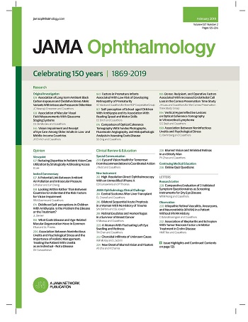
JAMA Ophthalmology, with a rich history of continuous publication since 1869, stands as a distinguished international, peer-reviewed journal dedicated to ophthalmology and visual science. In 2019, the journal proudly commemorated 150 years of uninterrupted service to the field. As a member of the esteemed JAMA Network, a consortium renowned for its peer-reviewed general medical and specialty publications, JAMA Ophthalmology upholds the highest standards of excellence in disseminating cutting-edge research and insights. Join us in celebrating our legacy and advancing the frontiers of ophthalmology and visual science.

 求助内容:
 应助结果提醒方式:


 京公网安备 11010802042870号


