
Assessing the technical capability of a room- and a gantry-mounted kV imaging device for intra-fractional fluoroscopy during stereotactic lung radiotherapy at a C-arm linear accelerator
IF 3.2
Q2 ONCOLOGY
Physics and Imaging in Radiation Oncology
Pub Date : 2026-05-01
Epub Date: 2026-05-02
DOI:10.1016/j.phro.2026.100988
引用次数: 0
Abstract
Background and Purpose:
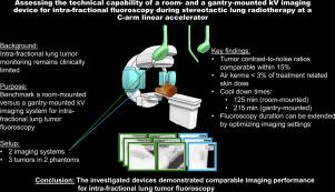
Monitoring lung tumors during stereotactic radiotherapy is important for managing breathing motion, yet the high-performance imaging modules on C-arm linear accelerators are mainly used for patient positioning. This study benchmarked a room-mounted against a gantry-mounted kilovolt (kV) imaging device to assess their potential for lung tumor fluoroscopy.
Materials and Methods:
The investigated systems were the ExacTrac Dynamic (ETD, Brainlab, Germany) and the X-ray Volumetric Imaging (XVI, Elekta, Sweden). Contrast-to-Noise Ratio (CNR) and entrance air kerma were measured for different imaging settings — tube voltage (70 kV to 130 kV), current (10 mA to 320 mA), exposure time (100 ms and 40 ms) — using two anthropomorphic thorax phantoms housing three tumors. Additionally, CNR change as a function of time, achievable fluoroscopy imaging duration based on X-ray generator and anode heat and X-ray tube cool-down were evaluated.
Results:
CNRs agreed within 15% for the two systems and changed less than 2% over the investigated fluoroscopy durations. Using default imaging settings and considering the average clinical duration of stereotactic lung treatments, entrance air kermas were 44 mGy (room-mounted) and 41 mGy (gantry-mounted). Achievable fluoroscopy durations with default settings were 87 s and 1350 s, respectively. Anode cool-down times were 125 min (room-mounted) and 215 min (gantry-mounted).
Conclusion:
The performance metrics of the room-mounted imaging system aligned with those of the gantry-mounted device with regard to CNR and air kerma. The room-mounted system showed a preferable cool-down behavior. However, adaptation of imaging settings is necessary to increase the fluoroscopy duration and avoid overheating.
评估在c臂直线加速器的立体定向肺放疗期间,房间式和龙门式kV成像装置用于分段透视的技术能力
背景与目的:立体定向放疗中监测肺肿瘤对控制呼吸运动很重要,而c臂直线加速器上的高性能成像模块主要用于患者定位。本研究对比了室内安装和龙门安装的千伏成像设备,以评估它们在肺肿瘤透视中的潜力。材料和方法:所研究的系统是ExacTrac Dynamic (ETD, Brainlab,德国)和x射线体积成像(XVI, Elekta,瑞典)。在不同的成像设置下——管电压(70千伏至130千伏),电流(10毫安至320毫安),曝光时间(100毫秒和40毫秒)——使用两个拟人胸腔幻象,容纳三个肿瘤,测量了对比噪声比(CNR)和入口空气密度。此外,还评估了CNR随时间的变化、基于x射线发生器、阳极加热和x射线管冷却的可实现的透视成像时间。结果:两种系统的cnr一致在15%以内,在调查的透视持续时间内变化不到2%。使用默认成像设置并考虑立体定向肺治疗的平均临床持续时间,入口空气角肿为44 mGy(室内安装)和41 mGy(龙门架安装)。默认设置下可实现的透视时间分别为87秒和1350秒。阳极冷却时间为125分钟(室内安装)和215分钟(龙门安装)。结论:室内成像系统与龙门式成像系统在CNR和空气质量方面的性能指标一致。房间安装系统显示出较好的冷却行为。然而,适应成像设置是必要的,以增加透视时间和避免过热。
本文章由计算机程序翻译,如有差异,请以英文原文为准。
求助全文
约1分钟内获得全文
求助全文
来源期刊
Physics and Imaging in Radiation Oncology
Physics and Astronomy-Radiation
CiteScore
5.30
自引率
18.90%
发文量
93
审稿时长
6 weeks
 求助内容:
求助内容: 应助结果提醒方式:

 京公网安备 11010802042870号


