
Automatic segmentation of the urethra and prostate zones with deep learning on T2-weighted magnetic resonance imaging

IF 3.2 Q2 ONCOLOGY
William Holmlund , Attila Simkó , Karin Söderkvist , Péter Palásti , Szilvia Tótin , Kamilla Kalmár , Zsófia Domoki , Zsuzsanna Fejes , Tamás Z. Kincses , Patrik Brynolfsson , Tufve Nyholm
{"title":"Automatic segmentation of the urethra and prostate zones with deep learning on T2-weighted magnetic resonance imaging","authors":"William Holmlund ,&nbsp;Attila Simkó ,&nbsp;Karin Söderkvist ,&nbsp;Péter Palásti ,&nbsp;Szilvia Tótin ,&nbsp;Kamilla Kalmár ,&nbsp;Zsófia Domoki ,&nbsp;Zsuzsanna Fejes ,&nbsp;Tamás Z. Kincses ,&nbsp;Patrik Brynolfsson ,&nbsp;Tufve Nyholm","doi":"10.1016/j.phro.2026.100964","DOIUrl":null,"url":null,"abstract":"<div><h3>Background and purpose</h3><div>Accurate segmentation of the urethra is crucial for safe focal dose escalated radiotherapy, while prostate zone identification is important for prostate cancer diagnosis. Manual delineations on magnetic resonance imaging (MRI) are labour-intensive and variable, and while deep learning offers promise in automating this process, no available solution currently exists. This study aimed to develop and evaluate a deep learning model for automatic segmentation of the urethra, prostate and all prostate zones and benchmark its performance against inter-reader variability and assess generalisability to external data from a different MRI vendor.</div></div><div><h3>Materials and methods</h3><div>The public datasets ProstateZones and PROSTATEx included 200 magnetic resonance images with manual delineations, with 160 used for training/validation and 40 with independent duplicate segmentations used as a test set. A nnU-Net deep learning model was evaluated on the unseen test set and externally validated on a dataset with 55 samples. Performance was assessed using Dice Similarity Coefficient (DSC), Surface DSC, percentile Symmetric Surface Distance, and Center Line Distance (CLD) metrics.</div></div><div><h3>Results</h3><div>The model outperformed the inter-reader variability on multiple structures, and notably on all metrics for the urethra, with median CLD values of 2.8 and 2.9 mm compared to 3.6 mm for inter-reader variability. External validation showed robust generalisability to a dataset collected from a different vendor.</div></div><div><h3>Conclusions</h3><div>This study demonstrated that a deep learning model can achieve expert-level performance in automated segmentation of the urethra, prostate, and prostate zones. Robust performance on external data highlighted potential as a decision support solution.</div></div>","PeriodicalId":36850,"journal":{"name":"Physics and Imaging in Radiation Oncology","volume":"38 ","pages":"Article 100964"},"PeriodicalIF":3.2000,"publicationDate":"2026-03-01","publicationTypes":"Journal Article","fieldsOfStudy":null,"isOpenAccess":false,"openAccessPdf":"","citationCount":"0","resultStr":null,"platform":"Semanticscholar","paperid":null,"PeriodicalName":"Physics and Imaging in Radiation Oncology","FirstCategoryId":"1085","ListUrlMain":"https://www.sciencedirect.com/science/article/pii/S2405631626000643","RegionNum":0,"RegionCategory":null,"ArticlePicture":[],"TitleCN":null,"AbstractTextCN":null,"PMCID":null,"EPubDate":"2026/4/7 0:00:00","PubModel":"Epub","JCR":"Q2","JCRName":"ONCOLOGY","Score":null,"Total":0}
引用次数: 0

Abstract

Background and purpose

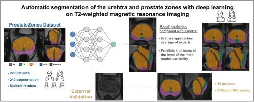
Accurate segmentation of the urethra is crucial for safe focal dose escalated radiotherapy, while prostate zone identification is important for prostate cancer diagnosis. Manual delineations on magnetic resonance imaging (MRI) are labour-intensive and variable, and while deep learning offers promise in automating this process, no available solution currently exists. This study aimed to develop and evaluate a deep learning model for automatic segmentation of the urethra, prostate and all prostate zones and benchmark its performance against inter-reader variability and assess generalisability to external data from a different MRI vendor.

Materials and methods

The public datasets ProstateZones and PROSTATEx included 200 magnetic resonance images with manual delineations, with 160 used for training/validation and 40 with independent duplicate segmentations used as a test set. A nnU-Net deep learning model was evaluated on the unseen test set and externally validated on a dataset with 55 samples. Performance was assessed using Dice Similarity Coefficient (DSC), Surface DSC, percentile Symmetric Surface Distance, and Center Line Distance (CLD) metrics.

Results

The model outperformed the inter-reader variability on multiple structures, and notably on all metrics for the urethra, with median CLD values of 2.8 and 2.9 mm compared to 3.6 mm for inter-reader variability. External validation showed robust generalisability to a dataset collected from a different vendor.

Conclusions

This study demonstrated that a deep learning model can achieve expert-level performance in automated segmentation of the urethra, prostate, and prostate zones. Robust performance on external data highlighted potential as a decision support solution.
Abstract Image

基于t2加权磁共振成像的深度学习自动分割尿道和前列腺区域
背景与目的准确的尿道分割对于安全的局灶剂量递增放射治疗至关重要,而前列腺分区的识别对于前列腺癌的诊断具有重要意义。磁共振成像(MRI)的人工描绘是劳动密集型和可变的,虽然深度学习在自动化这一过程中提供了希望,但目前还没有可用的解决方案。本研究旨在开发和评估一种用于自动分割尿道、前列腺和所有前列腺区域的深度学习模型,并根据阅读器间的可变性对其性能进行基准测试,并评估来自不同MRI供应商的外部数据的通用性。材料和方法公共数据集prostatzones和PROSTATEx包括200张手动描绘的磁共振图像,其中160张用于训练/验证,40张用于独立重复分割作为测试集。在未见的测试集上评估了nnU-Net深度学习模型,并在包含55个样本的数据集上进行了外部验证。使用骰子相似系数(DSC),表面DSC,百分位数对称表面距离和中心线距离(CLD)指标评估性能。结果该模型在多个结构上优于读写器间变异性,特别是在尿道的所有指标上,与读写器间变异性的3.6 mm相比,中位CLD值为2.8和2.9 mm。外部验证显示从不同供应商收集的数据集具有强大的通用性。本研究表明,深度学习模型可以在尿道、前列腺和前列腺区域的自动分割中达到专家级的性能。对外部数据的强大性能突出了作为决策支持解决方案的潜力。
本文章由计算机程序翻译,如有差异,请以英文原文为准。
求助全文
约1分钟内获得全文 求助全文
来源期刊
Physics and Imaging in Radiation Oncology
Physics and Imaging in Radiation Oncology Physics and Astronomy-Radiation
CiteScore
5.30
自引率
18.90%
发文量
93
审稿时长
6 weeks

×
引用
GB/T 7714-2015
复制
MLA
复制
APA
复制
导出至
BibTeX EndNote RefMan NoteFirst NoteExpress
×
提示
您的信息不完整,为了账户安全,请先补充。
现在去补充
×
提示
您因"违规操作"
具体请查看互助需知
我知道了
×
提示
确定
请完成安全验证×
copy
已复制链接
快去分享给好友吧!
我知道了
右上角分享
点击右上角分享
0
联系我们:info@booksci.cn Book学术提供免费学术资源搜索服务,方便国内外学者检索中英文文献。致力于提供最便捷和优质的服务体验。 Copyright © 2023 布克学术 All rights reserved.
京ICP备2023020795号-1
ghs 京公网安备 11010802042870号
Book学术文献互助
Book学术文献互助群
群 号:604180095
Book学术官方微信
小红书


