
Comparative analysis of artificial intelligence-based contouring of cardiac substructures on computed tomography scans for radiation therapy
IF 3.2
Q2 ONCOLOGY
Physics and Imaging in Radiation Oncology
Pub Date : 2026-03-01
Epub Date: 2026-02-21
DOI:10.1016/j.phro.2026.100935
引用次数: 0
Abstract
Background and purpose
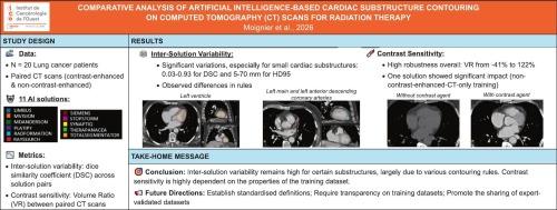
Artificial intelligence-based contouring tools enable assessment of radiation doses to cardiac substructures beyond mean heart dose. This study examined inter-solution variations in raw contours and the impact of non-contrast enhancement on contours for each solution.
Materials and methods
Contrast-enhanced (CE) and non-contrast-enhanced (NCE) breath-hold thoracic computed tomography (CT) scans, sequentially acquired during the same imaging session for twenty lung cancer patients, were used. Seven commercial, three open-source, and one in-house AI solutions were evaluated. On CE-CTs, solutions were compared using Dice Similarity Coefficient (DSC) and 95th percentile of Hausdorff distance (HD95) across each pair of solutions. Then, the effect of non-contrast enhancement on contours was assessed using volume ratios between NCE-CT and CE-CT for each solution.
Results
Typically, ten cardiac substructures were contoured by most of the solutions. For the whole heart, cardiac chambers and great vessels, the average median DSC was above 0.8 for 55 of the 123 structure-solution pairs (45%), and the average median HD95 was below 10 mm for 47 of the 123 structure-solution pairs (38%). For the coronary arteries, the average median DSC ranged between 0.03 and 0.50 and the average median HD95 ranged between 19 mm and 70 mm. Non-contrast enhancement influenced results variably; volume differences were below 10% for 84 of the 123 structure-solution pairs (68%).
Conclusions
Automatic contouring solutions exhibited inter-solution variability for cardiac substructures that may have clinical impact. Greater transparency and standardisation of models, ideally through international consensus and shared datasets, are essential.
放射治疗计算机断层扫描心脏亚结构轮廓的人工智能对比分析。
背景和目的:基于人工智能的轮廓工具可以评估心脏亚结构的辐射剂量超过平均心脏剂量。本研究考察了不同溶液间原始轮廓的变化,以及不同溶液下非对比度增强对轮廓的影响。材料和方法:使用对比增强(CE)和非对比增强(NCE)屏气胸部计算机断层扫描(CT)扫描,在同一成像过程中依次获得20例肺癌患者。评估了7个商业、3个开源和1个内部人工智能解决方案。在ce - ct上,使用骰子相似系数(DSC)和每对溶液的第95百分位豪斯多夫距离(HD95)对溶液进行比较。然后,使用NCE-CT和CE-CT对每种溶液的体积比评估非对比度增强对轮廓的影响。结果:大多数溶液均能勾勒出十个典型的心脏亚结构。对于整个心脏、心腔和大血管,123对结构-溶液对中有55对(45%)的平均中位DSC高于0.8,123对结构-溶液对中有47对(38%)的平均中位HD95低于10毫米。对于冠状动脉,平均中位DSC介于0.03至0.50之间,平均中位HD95介于19至70毫米之间。非对比增强对结果的影响是不同的;123个结构溶液对中有84个(68%)的体积差异低于10%。结论:自动轮廓溶液对心脏亚结构表现出溶液间的可变性,可能具有临床影响。提高模型的透明度和标准化至关重要,最好是通过国际共识和共享数据集实现。
本文章由计算机程序翻译,如有差异,请以英文原文为准。
求助全文
约1分钟内获得全文
求助全文
来源期刊
Physics and Imaging in Radiation Oncology
Physics and Astronomy-Radiation
CiteScore
5.30
自引率
18.90%
发文量
93
审稿时长
6 weeks
 求助内容:
求助内容: 应助结果提醒方式:

 京公网安备 11010802042870号


