Fusion and pure feature extraction framework for intraoperative hyperspectral of thyroid lesion
IF 11.8
1区 医学
Q1 COMPUTER SCIENCE, ARTIFICIAL INTELLIGENCE
引用次数: 0
Abstract
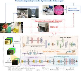
Thyroid cancer has remained one of the most prevalent endocrine malignancies. In routine surgery, thyroid cancer analysis involves two time-consuming steps: intraoperative frozen section preparation and manual microscopic examination. Recently, info-rich hyperspectral intelligence analysis has been studied, reducing subjective bias but only optimizing the intraoperative second step and the model complexity, ignoring the independent features that possess substance fingerprints. To bridge the gaps, we developed a hyperspectral recognition algorithm called PS4EM-SN for intraoperatively ex-vivo macro thyroid lesion, which comprised a pure spectral with pure spatial(SPS) learning framework and a spatial–spectral fusion embed mechanism(SSEM) coupled with cascade attention. The cascade attention mechanism, integrating Squeeze-and-Excitation (SE) and Non-Local (NOL) blocks, enhanced robustness to the outliers of SSEM and improved generalization. The experimental results were satisfactory in differentiating non-malignant and malignant regions with 93.91% average accuracy. Given its hyperspectral multifaceted performance, our method promises a digital solution for intraoperative thyroid diagnosis.
术中甲状腺病变高光谱融合与纯特征提取框架
甲状腺癌一直是最常见的内分泌恶性肿瘤之一。在常规手术中,甲状腺癌分析包括两个耗时的步骤:术中冰冻切片准备和人工显微镜检查。近年来,人们对信息丰富的高光谱智能分析进行了研究,减少了主观偏差,但只优化了术中第二步和模型复杂性,忽略了具有物质指纹的独立特征。为了弥补这一空白,我们开发了一种用于术中离体甲状腺大病变的高光谱识别算法PS4EM-SN,该算法由纯光谱与纯空间(SPS)学习框架和空间-光谱融合嵌入机制(SSEM)以及级联注意组成。层叠注意机制,整合了挤压-激励(SE)和非局部(NOL)块,增强了SSEM对异常值的鲁棒性,提高了泛化能力。实验结果对非恶性区和恶性区进行了满意的区分,平均准确率为93.91%。鉴于其高光谱多面性,我们的方法有望成为术中甲状腺诊断的数字解决方案。
本文章由计算机程序翻译,如有差异,请以英文原文为准。
求助全文
约1分钟内获得全文
求助全文
来源期刊

Medical image analysis
工程技术-工程:生物医学
CiteScore
22.10
自引率
6.40%
发文量
309
审稿时长
6.6 months
期刊介绍:
Medical Image Analysis serves as a platform for sharing new research findings in the realm of medical and biological image analysis, with a focus on applications of computer vision, virtual reality, and robotics to biomedical imaging challenges. The journal prioritizes the publication of high-quality, original papers contributing to the fundamental science of processing, analyzing, and utilizing medical and biological images. It welcomes approaches utilizing biomedical image datasets across all spatial scales, from molecular/cellular imaging to tissue/organ imaging.
 求助内容:
求助内容: 应助结果提醒方式:


