A new time-decay radiomics integrated network (TRINet) for breast cancer risk prediction
IF 11.8
1区 医学
Q1 COMPUTER SCIENCE, ARTIFICIAL INTELLIGENCE
引用次数: 0
Abstract
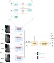
To facilitate early detection of breast cancer, there is a need to develop risk prediction schemes that can prescribe personalized screening mammography regimens for women. In this study, we propose a new deep learning architecture called TRINet that implements time-decay attention to focus on recent mammographic screenings, as current models do not account for the relevance of newer images. We integrate radiomic features with an Attention-based Multiple Instance Learning (AMIL) framework to weigh and combine multiple views for better risk estimation. In addition, we introduce a continual learning approach with a new label assignment strategy based on bilateral asymmetry to make the model more adaptable to asymmetrical cancer indicators. Finally, we add a time-embedded additive hazard layer to perform dynamic, multi-year risk forecasting based on individualized screening intervals. We used two public datasets, namely 8528 patients from the American EMBED dataset and 8723 patients from the Swedish CSAW dataset in our experiments. Evaluation results on the EMBED test set show that our approach performs comparably with state-of-the-art models, achieving AUC scores of 0.851, 0.811, 0.796, 0.793, and 0.789 across 1-, 2-, to 5-year intervals, respectively. Our results underscore the importance of integrating temporal attention, radiomic features, time embeddings, bilateral asymmetry, and continual learning strategies, providing a more adaptive and precise tool for breast cancer risk prediction.
一种新的时间衰减放射组学集成网络(TRINet)用于乳腺癌风险预测
为了促进乳腺癌的早期发现,有必要制定风险预测方案,为妇女开出个性化的乳房x光检查方案。在这项研究中,我们提出了一种新的深度学习架构,称为TRINet,它实现了时间衰减关注,专注于最近的乳房x光检查,因为当前的模型没有考虑到新图像的相关性。我们将放射学特征与基于注意力的多实例学习(AMIL)框架相结合,以权衡和组合多个视图,从而更好地进行风险评估。此外,我们引入了一种基于双边不对称的新的标签分配策略的持续学习方法,使模型更能适应不对称的癌症指标。最后,我们添加了一个时间嵌入的附加危害层,以基于个性化筛查间隔进行动态的多年风险预测。在我们的实验中,我们使用了两个公共数据集,即来自美国EMBED数据集的8528例患者和来自瑞典CSAW数据集的8723例患者。在EMBED测试集上的评估结果表明,我们的方法与最先进的模型表现相当,在1年、2年和5年的时间间隔内,AUC得分分别为0.851、0.811、0.796、0.793和0.789。我们的研究结果强调了整合时间注意力、放射学特征、时间嵌入、双侧不对称和持续学习策略的重要性,为乳腺癌风险预测提供了更具适应性和准确性的工具。
本文章由计算机程序翻译,如有差异,请以英文原文为准。
求助全文
约1分钟内获得全文
求助全文
来源期刊

Medical image analysis
工程技术-工程:生物医学
CiteScore
22.10
自引率
6.40%
发文量
309
审稿时长
6.6 months
期刊介绍:
Medical Image Analysis serves as a platform for sharing new research findings in the realm of medical and biological image analysis, with a focus on applications of computer vision, virtual reality, and robotics to biomedical imaging challenges. The journal prioritizes the publication of high-quality, original papers contributing to the fundamental science of processing, analyzing, and utilizing medical and biological images. It welcomes approaches utilizing biomedical image datasets across all spatial scales, from molecular/cellular imaging to tissue/organ imaging.
 求助内容:
求助内容: 应助结果提醒方式:


