{"title":"Mitigation of T<sub>1</sub> impact for unbiased tumor magnetic resonance amide proton transfer imaging at 3T.","authors":"Zhou Liu, Qian Yang, Haizhou Liu, Honghong Luo, Yijia Zheng, Dehong Luo, Yin Wu","doi":"10.1093/radadv/umaf011","DOIUrl":null,"url":null,"abstract":"<p><strong>Background: </strong>Amide proton transfer (APT), a specific type of chemical exchange saturation transfer (CEST) MRI, has proved valuable in tumor diagnosis and characterization by detecting mobile protein/peptides in cancerous tissues. However, T<sub>1</sub> confounds CEST measurements, leading to reduced specificity to amides and potential misinterpretation of APT imaging.</p><p><strong>Purpose: </strong>The study aimed to investigate the feasibility of the quasi-steady-state (QUASS)-based apparent exchange-dependent relaxation (AREX) analysis in correcting T<sub>1</sub> for unbiased tumor APT MRI at 3T.</p><p><strong>Materials and methods: </strong>CEST MRI experiments were conducted on an egg white phantom and on prospectively enrolled brain tumor patients with T<sub>1</sub> values modulated by gadolinium (Gd). QUASS algorithm was employed to reconstruct steady-state Z spectra. Conventional T<sub>1</sub>-uncorrected CEST effect was quantified with a multipool Lorentzian function from QUASS Z spectra. The non-QUASS AREX and QUASS-based AREX with T<sub>1</sub> correction were calculated from the inverse of non-QUASS and QUASS Z spectra, respectively. The student's <i>t</i>-test and Bland-Altman plots were performed to assess the statistical difference and consistency between pre- and post-Gd measurements.</p><p><strong>Results: </strong>In the phantom study, vials with different T<sub>1</sub> values showed conspicuous discrepancy on the conventional uncorrected APT and non-QUASS AREX maps, but comparable contrast on the QUASS-based AREX map. In the human study, 13 patients were enrolled. The contralateral normal-appearing white matter exhibited no substantial change in T<sub>1</sub> and similar CEST effect between uncorrected APT, non-QUASS AREX, and QUASS-based AREX pre- and post-Gd (all <i>P</i> > .05). However, the tumor regions showed significantly reduced T<sub>1</sub> post-Gd that altered the CEST measurements obtained from uncorrected APT and non-QUASS AREX (both <i>P</i> < .001). In comparison, QUASS-based AREX measurements were in excellent agreement between pre- and post-Gd (<i>P</i> = .19).</p><p><strong>Conclusion: </strong>QUASS-based AREX analysis can effectively correct T<sub>1</sub> contamination in CEST measurements, facilitating unbiased tumor APT MRI at 3T.</p>","PeriodicalId":519940,"journal":{"name":"Radiology advances","volume":"2 2","pages":"umaf011"},"PeriodicalIF":0.0000,"publicationDate":"2025-03-07","publicationTypes":"Journal Article","fieldsOfStudy":null,"isOpenAccess":false,"openAccessPdf":"https://www.ncbi.nlm.nih.gov/pmc/articles/PMC12429276/pdf/","citationCount":"0","resultStr":null,"platform":"Semanticscholar","paperid":null,"PeriodicalName":"Radiology advances","FirstCategoryId":"1085","ListUrlMain":"https://doi.org/10.1093/radadv/umaf011","RegionNum":0,"RegionCategory":null,"ArticlePicture":[],"TitleCN":null,"AbstractTextCN":null,"PMCID":null,"EPubDate":"2025/3/1 0:00:00","PubModel":"eCollection","JCR":"","JCRName":"","Score":null,"Total":0}
引用次数: 0
Abstract
Background: Amide proton transfer (APT), a specific type of chemical exchange saturation transfer (CEST) MRI, has proved valuable in tumor diagnosis and characterization by detecting mobile protein/peptides in cancerous tissues. However, T1 confounds CEST measurements, leading to reduced specificity to amides and potential misinterpretation of APT imaging.
Purpose: The study aimed to investigate the feasibility of the quasi-steady-state (QUASS)-based apparent exchange-dependent relaxation (AREX) analysis in correcting T1 for unbiased tumor APT MRI at 3T.
Materials and methods: CEST MRI experiments were conducted on an egg white phantom and on prospectively enrolled brain tumor patients with T1 values modulated by gadolinium (Gd). QUASS algorithm was employed to reconstruct steady-state Z spectra. Conventional T1-uncorrected CEST effect was quantified with a multipool Lorentzian function from QUASS Z spectra. The non-QUASS AREX and QUASS-based AREX with T1 correction were calculated from the inverse of non-QUASS and QUASS Z spectra, respectively. The student's t-test and Bland-Altman plots were performed to assess the statistical difference and consistency between pre- and post-Gd measurements.
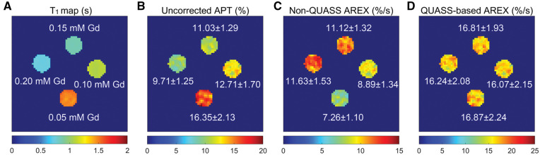
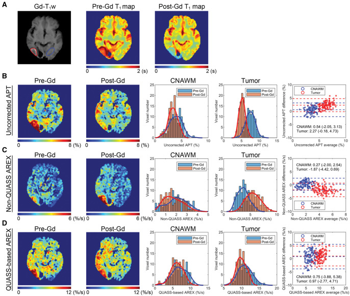
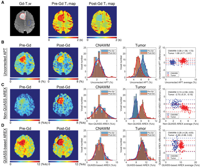
Results: In the phantom study, vials with different T1 values showed conspicuous discrepancy on the conventional uncorrected APT and non-QUASS AREX maps, but comparable contrast on the QUASS-based AREX map. In the human study, 13 patients were enrolled. The contralateral normal-appearing white matter exhibited no substantial change in T1 and similar CEST effect between uncorrected APT, non-QUASS AREX, and QUASS-based AREX pre- and post-Gd (all P > .05). However, the tumor regions showed significantly reduced T1 post-Gd that altered the CEST measurements obtained from uncorrected APT and non-QUASS AREX (both P < .001). In comparison, QUASS-based AREX measurements were in excellent agreement between pre- and post-Gd (P = .19).
Conclusion: QUASS-based AREX analysis can effectively correct T1 contamination in CEST measurements, facilitating unbiased tumor APT MRI at 3T.

 求助内容:
求助内容: 应助结果提醒方式:


