Mingwei Xie, Haonan Wang, Zehong Yang, Ming Gao, Guangzi Shi, Xingnan Liao, Zhongqiang Luo, Xiaomeng Li, Jun Shen
{"title":"Artificial intelligence model for automatic 3-dimensional reconstruction of ossicular chain and bony labyrinth from high-resolution CT.","authors":"Mingwei Xie, Haonan Wang, Zehong Yang, Ming Gao, Guangzi Shi, Xingnan Liao, Zhongqiang Luo, Xiaomeng Li, Jun Shen","doi":"10.1093/radadv/umaf004","DOIUrl":null,"url":null,"abstract":"<p><strong>Background: </strong>Three-dimensional (3D) reconstruction of ossicular chain and bony labyrinth based on temporal bone high-resolution CT (HRCT) is useful for diagnosis and treatment guidance of middle and inner ear diseases. However, these structures are small and irregular, making manual reconstruction time-consuming.</p><p><strong>Purpose: </strong>To develop and validate an artificial intelligence (AI) model based on semisupervised learning for automated 3D reconstruction of ossicular chain and bony labyrinth on HRCT images.</p><p><strong>Methods: </strong>HRCT images from 304 ears of 152 consecutive patients retrospectively collected from a single center were randomly divided into training (246 ears), validation (28 ears), and internal test (30 ears) cohorts for model development. A novel semisupervised ear bone segmentation framework was used to train the AI model, and its performance was evaluated by Dice similarity coefficients. The trained algorithm was applied to a temporally independent test dataset of 30 ears of 15 patients from the same center for comparison with manual 3D reconstruction for processing time, target volume, and visual assessment of segmentation.</p><p><strong>Results: </strong>The AI model demonstrated a Dice score of 0.948 (95% CI, 0.940-0.955) for the internal and 0.979 (95% CI, 0.973-0.986) for the temporally independent test sets. In the latter dataset, the AI model required 2% or less processing time of manual 3D reconstruction for each ear (17.7 seconds ± 10.1 vs 1080.5 seconds ± 149.8; <i>P</i> < .001) and had an accuracy comparable to human experts in the volume and visual assessment of segmentation targets (<i>P</i> = .237-1.000). In a subgroup analysis, the model achieved accurate segmentation (Dice scores of 0.98-0.99) across various diseases (eg, otitis media, mastoiditis, otosclerosis, middle and inner ear malformations, Ménière disease).</p><p><strong>Conclusion: </strong>The AI model enables robust, efficient and accurate 3D reconstruction for the small structures such as ossicular chain and bony labyrinth on HRCT images.</p>","PeriodicalId":519940,"journal":{"name":"Radiology advances","volume":"2 1","pages":"umaf004"},"PeriodicalIF":0.0000,"publicationDate":"2025-01-28","publicationTypes":"Journal Article","fieldsOfStudy":null,"isOpenAccess":false,"openAccessPdf":"https://www.ncbi.nlm.nih.gov/pmc/articles/PMC12429245/pdf/","citationCount":"0","resultStr":null,"platform":"Semanticscholar","paperid":null,"PeriodicalName":"Radiology advances","FirstCategoryId":"1085","ListUrlMain":"https://doi.org/10.1093/radadv/umaf004","RegionNum":0,"RegionCategory":null,"ArticlePicture":[],"TitleCN":null,"AbstractTextCN":null,"PMCID":null,"EPubDate":"2025/1/1 0:00:00","PubModel":"eCollection","JCR":"","JCRName":"","Score":null,"Total":0}
引用次数: 0
Abstract
Background: Three-dimensional (3D) reconstruction of ossicular chain and bony labyrinth based on temporal bone high-resolution CT (HRCT) is useful for diagnosis and treatment guidance of middle and inner ear diseases. However, these structures are small and irregular, making manual reconstruction time-consuming.
Purpose: To develop and validate an artificial intelligence (AI) model based on semisupervised learning for automated 3D reconstruction of ossicular chain and bony labyrinth on HRCT images.
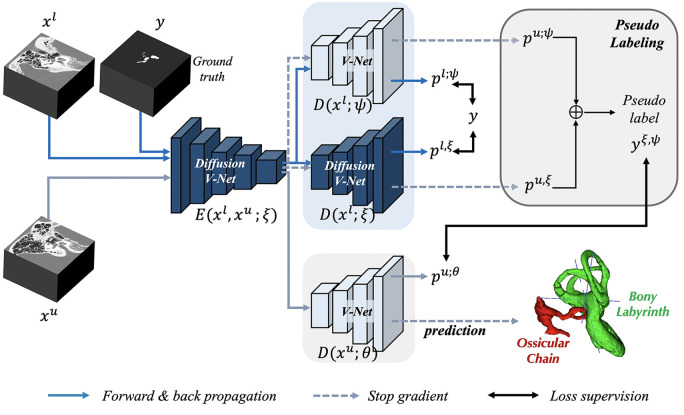
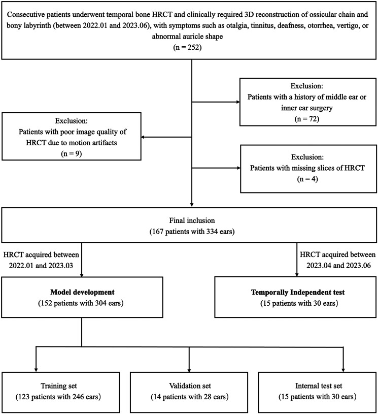
Methods: HRCT images from 304 ears of 152 consecutive patients retrospectively collected from a single center were randomly divided into training (246 ears), validation (28 ears), and internal test (30 ears) cohorts for model development. A novel semisupervised ear bone segmentation framework was used to train the AI model, and its performance was evaluated by Dice similarity coefficients. The trained algorithm was applied to a temporally independent test dataset of 30 ears of 15 patients from the same center for comparison with manual 3D reconstruction for processing time, target volume, and visual assessment of segmentation.
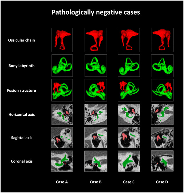
Results: The AI model demonstrated a Dice score of 0.948 (95% CI, 0.940-0.955) for the internal and 0.979 (95% CI, 0.973-0.986) for the temporally independent test sets. In the latter dataset, the AI model required 2% or less processing time of manual 3D reconstruction for each ear (17.7 seconds ± 10.1 vs 1080.5 seconds ± 149.8; P < .001) and had an accuracy comparable to human experts in the volume and visual assessment of segmentation targets (P = .237-1.000). In a subgroup analysis, the model achieved accurate segmentation (Dice scores of 0.98-0.99) across various diseases (eg, otitis media, mastoiditis, otosclerosis, middle and inner ear malformations, Ménière disease).
Conclusion: The AI model enables robust, efficient and accurate 3D reconstruction for the small structures such as ossicular chain and bony labyrinth on HRCT images.

 求助内容:
求助内容: 应助结果提醒方式:


