Laura Onac, Lorand Dobai, Andrei Mouraviev, Mihai A Badila, Denise Yap, Samantha Kennedy, Annie Nguyen, Emi Gal, Daniel K Sodickson
{"title":"An image-domain deep-learning denoising technique for accelerated parallel brain MRI: prospective clinical evaluation.","authors":"Laura Onac, Lorand Dobai, Andrei Mouraviev, Mihai A Badila, Denise Yap, Samantha Kennedy, Annie Nguyen, Emi Gal, Daniel K Sodickson","doi":"10.1093/radadv/umae022","DOIUrl":null,"url":null,"abstract":"<p><strong>Background: </strong>Parallel imaging can accelerate MRI acquisitions, but excessive accelerations can introduce amplified noise and aliasing artifacts.</p><p><strong>Purpose: </strong>To evaluate a vendor-agnostic AI-based approach to remove image degradation artifacts in highly accelerated MRI scans, improving image quality and reducing scan time.</p><p><strong>Materials and methods: </strong>Training was performed by retrospectively degrading standard accelerated images. Evaluation was performed with both retrospective and prospectively-collected highly accelerated images. Retrospective data were taken from ∼2000 MRI studies obtained between August 2016 and October 2022, and prospective data were collected in >200 subjects between June and November 2022, using scanners from multiple vendors and locations. Scan time data were collected from prospective studies and used to compute time savings per sequence and per protocol for each vendor. Images were evaluated qualitatively by 5 board-certified radiologists and quantitatively by assessing noise, contrast, and spatial resolution. Paired Wilcoxon signed-rank tests were used to compare model outputs to model inputs and low-acceleration images.</p><p><strong>Results: </strong>Images from 101 adults from 5 sites and 6 scanner models from different vendors were enrolled. 89% of imaged subjects had noteworthy imaging features or pathology. Model outputs were rated superior to model inputs (<i>P < .</i>001) and most were either non-inferior (<i>P</i> <sub>inf</sub> <i> > </i>.05) or superior (<i>P</i> <sub>sup</sub> <i> < </i>.05) to baseline images in qualitative metrics of image quality and feature visibility. Quantitative evaluation of signal-to-noise ratio and contrast-to-noise ratio improved for model outputs compared to inputs (<i>P < .</i>001) or baseline images (<i>P < .</i>005). Apparent resolution measured using the full width at half maximum or minimum was either enhanced (<i>P</i> <sub>sup</sub> <i> < </i>.05) or preserved (non-superior <i>P</i> <sub>sup</sub> <i> > </i>.05 and non-inferior <i>P</i> <sub>inf</sub> <i> > </i>.05). The scan time was reduced by an average of 29% (19%-41% per sequence).</p><p><strong>Conclusion: </strong>This vendor-agnostic AI-based method achieved robust scan time savings without loss of image quality, potentially allowing for reduced cost and improved patient experience.</p>","PeriodicalId":519940,"journal":{"name":"Radiology advances","volume":"1 3","pages":"umae022"},"PeriodicalIF":0.0000,"publicationDate":"2024-08-14","publicationTypes":"Journal Article","fieldsOfStudy":null,"isOpenAccess":false,"openAccessPdf":"https://www.ncbi.nlm.nih.gov/pmc/articles/PMC12481681/pdf/","citationCount":"0","resultStr":null,"platform":"Semanticscholar","paperid":null,"PeriodicalName":"Radiology advances","FirstCategoryId":"1085","ListUrlMain":"https://doi.org/10.1093/radadv/umae022","RegionNum":0,"RegionCategory":null,"ArticlePicture":[],"TitleCN":null,"AbstractTextCN":null,"PMCID":null,"EPubDate":"2024/9/1 0:00:00","PubModel":"eCollection","JCR":"","JCRName":"","Score":null,"Total":0}
引用次数: 0
Abstract
Background: Parallel imaging can accelerate MRI acquisitions, but excessive accelerations can introduce amplified noise and aliasing artifacts.
Purpose: To evaluate a vendor-agnostic AI-based approach to remove image degradation artifacts in highly accelerated MRI scans, improving image quality and reducing scan time.
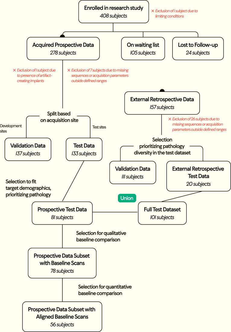
Materials and methods: Training was performed by retrospectively degrading standard accelerated images. Evaluation was performed with both retrospective and prospectively-collected highly accelerated images. Retrospective data were taken from ∼2000 MRI studies obtained between August 2016 and October 2022, and prospective data were collected in >200 subjects between June and November 2022, using scanners from multiple vendors and locations. Scan time data were collected from prospective studies and used to compute time savings per sequence and per protocol for each vendor. Images were evaluated qualitatively by 5 board-certified radiologists and quantitatively by assessing noise, contrast, and spatial resolution. Paired Wilcoxon signed-rank tests were used to compare model outputs to model inputs and low-acceleration images.
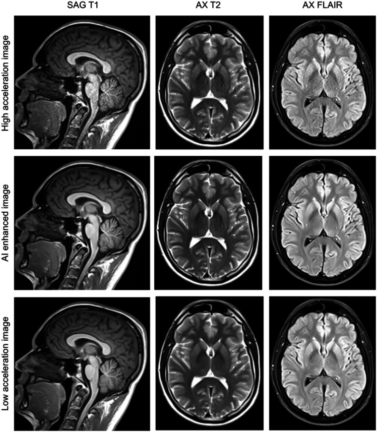
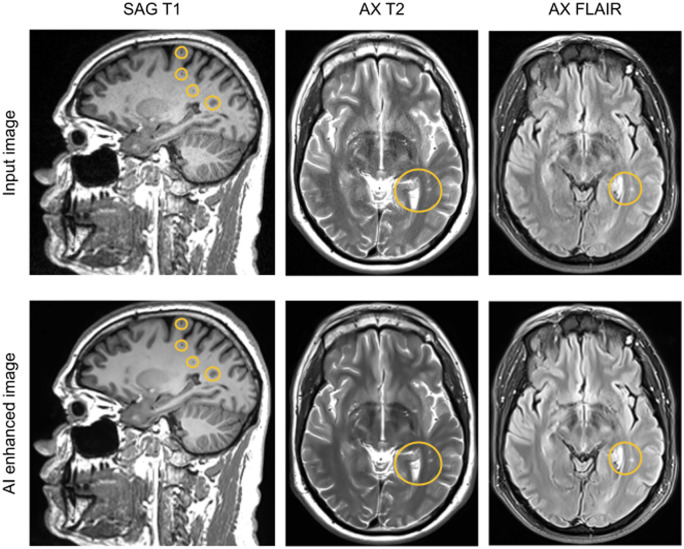
Results: Images from 101 adults from 5 sites and 6 scanner models from different vendors were enrolled. 89% of imaged subjects had noteworthy imaging features or pathology. Model outputs were rated superior to model inputs (P < .001) and most were either non-inferior (Pinf > .05) or superior (Psup < .05) to baseline images in qualitative metrics of image quality and feature visibility. Quantitative evaluation of signal-to-noise ratio and contrast-to-noise ratio improved for model outputs compared to inputs (P < .001) or baseline images (P < .005). Apparent resolution measured using the full width at half maximum or minimum was either enhanced (Psup < .05) or preserved (non-superior Psup > .05 and non-inferior Pinf > .05). The scan time was reduced by an average of 29% (19%-41% per sequence).
Conclusion: This vendor-agnostic AI-based method achieved robust scan time savings without loss of image quality, potentially allowing for reduced cost and improved patient experience.

 求助内容:
求助内容: 应助结果提醒方式:


