Rolf Reiter, Pinkas Mürdel, Florian N Loch, Mehrgan Shahryari, Rebecca Strehle, Christian Bayerl, Jürgen Braun, Ingolf Sack, Patrick Asbach, David Kaufmann
{"title":"Intervertebral disc degeneration of the lumbar spine assessed in vivo with 3T magnetic resonance tomoelastography.","authors":"Rolf Reiter, Pinkas Mürdel, Florian N Loch, Mehrgan Shahryari, Rebecca Strehle, Christian Bayerl, Jürgen Braun, Ingolf Sack, Patrick Asbach, David Kaufmann","doi":"10.1093/radadv/umaf013","DOIUrl":null,"url":null,"abstract":"<p><strong>Introduction: </strong>Assessment of intervertebral disc (IVD) degeneration on conventional magnetic resonance imaging (MRI) is limited by large inter-reader variability and lack of stratification in clinical trials and their assessment of treatment responses. Therefore, we aimed to introduce and diagnostically validate multifrequency magnetic resonance elastography (MRE) with tomoelastography processing for the assessment of lumbar IVD degeneration in healthy volunteers and patients with low back pain.</p><p><strong>Methods: </strong>In this prospective single-center study, 60 participants (30 volunteers without low back pain and 30 patients with low back pain, 41 ± 17 years, body mass index 23.9 ± 3.7 kg/m<sup>2</sup>, 25 women) underwent multifrequency MRE using vibration frequencies from 40 to 70 Hz using a custom-built MRE setup in a 3T MRI scanner (Magnetom Skyra, software version XA30, Siemens Healthineers, Erlangen, Germany). Maps of shear wave speed (SWS in m/s) and loss angle (<i>φ</i> in rad), representing stiffness and viscous properties, respectively, were generated using tomoelastography data processing. The Pfirrmann score was used as reference standard to assess lumbar IVD degeneration on sagittal T2-weighted images. Inter-reader agreement (3 readers) and repeatability were assessed using the intraclass correlation coefficient (ICC).</p><p><strong>Results: </strong>Area under the receiver operating characteristic curve (AUC) analysis showed good diagnostic performance for detecting IVD degeneration (Pfirrmann score I/II/III/IV/V with <i>n</i> = 7/18/9/18/9, respectively) based on SWS (AUC: ≥II: 0.83, ≥III: 0.91, ≥IV: 0.96, V: 0.97) and <i>φ</i> (AUC: ≥II: 0.88, ≥III: 0.93, ≥IV: 0.98, V: 0.95). Good and excellent inter-reader agreements were found for Pfirrmann score (ICC = 0.87), SWS (ICC = 0.87), and <i>φ</i> (ICC = 0.92), respectively. Good repeatability was demonstrated for SWS (ICC = 0.88) and <i>φ</i> (ICC = 0.88).</p><p><strong>Discussion: </strong>Multifrequency MRE with tomoelastography processing allows effective IVD assessment and shows promise as a quantitative clinical imaging modality for assessing IVD degeneration.</p>","PeriodicalId":519940,"journal":{"name":"Radiology advances","volume":"2 3","pages":"umaf013"},"PeriodicalIF":0.0000,"publicationDate":"2025-05-08","publicationTypes":"Journal Article","fieldsOfStudy":null,"isOpenAccess":false,"openAccessPdf":"https://www.ncbi.nlm.nih.gov/pmc/articles/PMC12429253/pdf/","citationCount":"0","resultStr":null,"platform":"Semanticscholar","paperid":null,"PeriodicalName":"Radiology advances","FirstCategoryId":"1085","ListUrlMain":"https://doi.org/10.1093/radadv/umaf013","RegionNum":0,"RegionCategory":null,"ArticlePicture":[],"TitleCN":null,"AbstractTextCN":null,"PMCID":null,"EPubDate":"2025/5/1 0:00:00","PubModel":"eCollection","JCR":"","JCRName":"","Score":null,"Total":0}
引用次数: 0
Abstract
Introduction: Assessment of intervertebral disc (IVD) degeneration on conventional magnetic resonance imaging (MRI) is limited by large inter-reader variability and lack of stratification in clinical trials and their assessment of treatment responses. Therefore, we aimed to introduce and diagnostically validate multifrequency magnetic resonance elastography (MRE) with tomoelastography processing for the assessment of lumbar IVD degeneration in healthy volunteers and patients with low back pain.
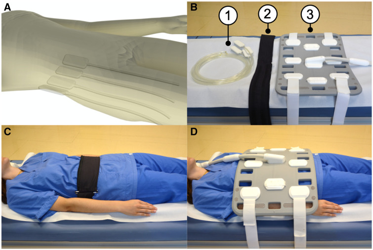
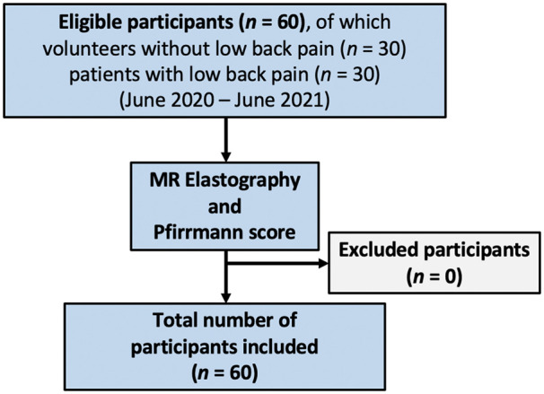
Methods: In this prospective single-center study, 60 participants (30 volunteers without low back pain and 30 patients with low back pain, 41 ± 17 years, body mass index 23.9 ± 3.7 kg/m2, 25 women) underwent multifrequency MRE using vibration frequencies from 40 to 70 Hz using a custom-built MRE setup in a 3T MRI scanner (Magnetom Skyra, software version XA30, Siemens Healthineers, Erlangen, Germany). Maps of shear wave speed (SWS in m/s) and loss angle (φ in rad), representing stiffness and viscous properties, respectively, were generated using tomoelastography data processing. The Pfirrmann score was used as reference standard to assess lumbar IVD degeneration on sagittal T2-weighted images. Inter-reader agreement (3 readers) and repeatability were assessed using the intraclass correlation coefficient (ICC).
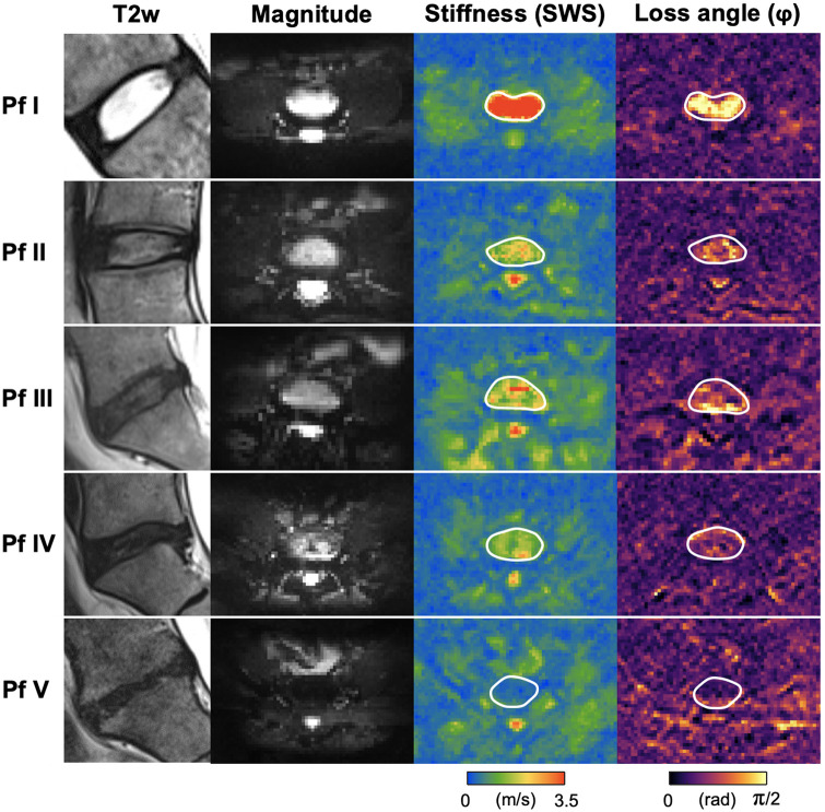
Results: Area under the receiver operating characteristic curve (AUC) analysis showed good diagnostic performance for detecting IVD degeneration (Pfirrmann score I/II/III/IV/V with n = 7/18/9/18/9, respectively) based on SWS (AUC: ≥II: 0.83, ≥III: 0.91, ≥IV: 0.96, V: 0.97) and φ (AUC: ≥II: 0.88, ≥III: 0.93, ≥IV: 0.98, V: 0.95). Good and excellent inter-reader agreements were found for Pfirrmann score (ICC = 0.87), SWS (ICC = 0.87), and φ (ICC = 0.92), respectively. Good repeatability was demonstrated for SWS (ICC = 0.88) and φ (ICC = 0.88).
Discussion: Multifrequency MRE with tomoelastography processing allows effective IVD assessment and shows promise as a quantitative clinical imaging modality for assessing IVD degeneration.

 求助内容:
求助内容: 应助结果提醒方式:


