An Interpretable Radiomics-Based Model Using Susceptibility-Weighted Imaging for Non-Invasive Prediction of Tertiary Lymphoid Structures in Hepatocellular Carcinoma.
Lizhen Liu, Fen Gao, Yiman Li, Jie Cheng, Huarong Zhang, Ping Cai, Wei Chen, Xiaoming Li
{"title":"An Interpretable Radiomics-Based Model Using Susceptibility-Weighted Imaging for Non-Invasive Prediction of Tertiary Lymphoid Structures in Hepatocellular Carcinoma.","authors":"Lizhen Liu, Fen Gao, Yiman Li, Jie Cheng, Huarong Zhang, Ping Cai, Wei Chen, Xiaoming Li","doi":"10.2147/JHC.S551462","DOIUrl":null,"url":null,"abstract":"<p><strong>Background: </strong>Intratumoral tertiary lymphoid structures (iTLSs) are associated with favorable prognosis and immunotherapy response in hepatocellular carcinoma (HCC). This study aimed to develop an interpretable susceptibility-weighted imaging (SWI)-based radiomics model to non-invasively predict iTLSs in HCC.</p><p><strong>Materials and methods: </strong>A retrospective cohort of 477 HCC patients undergoing preoperative SWI was used (training: 290; validation: 125; independent validation: 62). Radiomics models were constructed using five machine learning algorithms: logistic regression, random forest (RF), support vector machine, extreme gradient boosting, and K-nearest neighbors. Model performance was evaluated using the area under the ROC curve (AUC), model interpretability was examined using shapley additive explanations (SHAP), and survival analyses were performed to assess clinical relevance.</p><p><strong>Results: </strong>In the independent validation cohort, the RF algorithm was identified as the optimal classifier, with an AUC of 0.771 (95% CI: 0.641-0.883), sensitivity of 78.6%, and specificity of 67.6%. It significantly outperformed the radiological model (p = 0.046), and showed comparable performance with the hybrid model in predicting iTLSs positivity (iTLSs+) (p > 0.05). SHAP analysis showed that radiomics features (logarithm_firstorder_Minimum and exponential_glszm_ZoneEntropy) were significant predictors of iTLSs+. Kaplan-Meier analysis demonstrated improved time-to-recurrence (TTR) in the iTLSs+ predictor group compared to the iTLSs-negativity (iTLSs-) predictor group (p < 0.05). Furthermore, patients in the iTLSs+ predictor group receiving tyrosine kinase inhibitors combined with immune checkpoint inhibitors (TKI-ICI) therapy exhibited significantly extended TTR (p < 0.05), while no benefit was observed in the iTLSs- predictor group.</p><p><strong>Conclusion: </strong>The SWI-based radiomics model provided a non-invasive tool for predicting iTLSs+ in HCC and identifying patients who might benefit from TKI-ICI therapy, and it showed potential for future integration into clinical decision-making workflows.</p>","PeriodicalId":15906,"journal":{"name":"Journal of Hepatocellular Carcinoma","volume":"12 ","pages":"2197-2211"},"PeriodicalIF":3.4000,"publicationDate":"2025-09-30","publicationTypes":"Journal Article","fieldsOfStudy":null,"isOpenAccess":false,"openAccessPdf":"https://www.ncbi.nlm.nih.gov/pmc/articles/PMC12495958/pdf/","citationCount":"0","resultStr":null,"platform":"Semanticscholar","paperid":null,"PeriodicalName":"Journal of Hepatocellular Carcinoma","FirstCategoryId":"3","ListUrlMain":"https://doi.org/10.2147/JHC.S551462","RegionNum":3,"RegionCategory":"医学","ArticlePicture":[],"TitleCN":null,"AbstractTextCN":null,"PMCID":null,"EPubDate":"2025/1/1 0:00:00","PubModel":"eCollection","JCR":"Q2","JCRName":"ONCOLOGY","Score":null,"Total":0}
引用次数: 0
Abstract
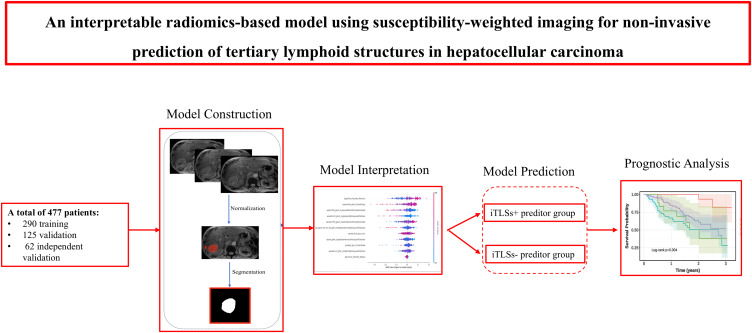
Background: Intratumoral tertiary lymphoid structures (iTLSs) are associated with favorable prognosis and immunotherapy response in hepatocellular carcinoma (HCC). This study aimed to develop an interpretable susceptibility-weighted imaging (SWI)-based radiomics model to non-invasively predict iTLSs in HCC.
Materials and methods: A retrospective cohort of 477 HCC patients undergoing preoperative SWI was used (training: 290; validation: 125; independent validation: 62). Radiomics models were constructed using five machine learning algorithms: logistic regression, random forest (RF), support vector machine, extreme gradient boosting, and K-nearest neighbors. Model performance was evaluated using the area under the ROC curve (AUC), model interpretability was examined using shapley additive explanations (SHAP), and survival analyses were performed to assess clinical relevance.
Results: In the independent validation cohort, the RF algorithm was identified as the optimal classifier, with an AUC of 0.771 (95% CI: 0.641-0.883), sensitivity of 78.6%, and specificity of 67.6%. It significantly outperformed the radiological model (p = 0.046), and showed comparable performance with the hybrid model in predicting iTLSs positivity (iTLSs+) (p > 0.05). SHAP analysis showed that radiomics features (logarithm_firstorder_Minimum and exponential_glszm_ZoneEntropy) were significant predictors of iTLSs+. Kaplan-Meier analysis demonstrated improved time-to-recurrence (TTR) in the iTLSs+ predictor group compared to the iTLSs-negativity (iTLSs-) predictor group (p < 0.05). Furthermore, patients in the iTLSs+ predictor group receiving tyrosine kinase inhibitors combined with immune checkpoint inhibitors (TKI-ICI) therapy exhibited significantly extended TTR (p < 0.05), while no benefit was observed in the iTLSs- predictor group.
Conclusion: The SWI-based radiomics model provided a non-invasive tool for predicting iTLSs+ in HCC and identifying patients who might benefit from TKI-ICI therapy, and it showed potential for future integration into clinical decision-making workflows.
 求助内容:
求助内容: 应助结果提醒方式:


