Leveraging dixon-based magnetic resonance imaging for pelvic bone marrow imaging in radiotherapy
IF 3.3
Q2 ONCOLOGY
引用次数: 0
Abstract
Background and purpose
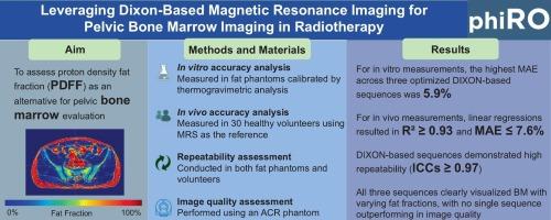
Pelvic radiotherapy-induced bone marrow (BM) damage adversely affects patient prognosis. Progress in BM-sparing radiotherapy is limited by the lack of standardized BM quantification and the inherent constraints of magnetic resonance spectroscopy (MRS), the current gold standard for BM magnetic resonance imaging (MRI). Proton density fat fraction (PDFF), derived from DIXON-based MRI, has emerged as an imaging biomarker for detecting BM changes. This study evaluated the potential of DIXON-based MRI in pelvic BM for radiotherapy.
Materials and methods
Three existing DIXON-based techniques were optimized and compared to establish clinical protocols. In vitro measurements were performed using fat phantoms calibrated against thermogravimetric analysis, while in vivo measurements were based on data from 30 volunteers with MRS serving as the reference standard. Quantitative accuracy was assessed using mean absolute error (MAE), repeatability via intra-class correlation coefficients (ICCs), and image quality using an ACR phantom.
Results
Comprehensive evaluation identified optimal parameters for each DIXON-based sequence. For in vitro measurements, the MAE for MRS was 3.5 % and the highest MAE across three optimized DIXON-based sequences was 5.9 %. For in vivo measurements, linear regressions between MRS and each of the optimized DIXON-based sequence resulted in R2 ≥ 0.93 and MAE ≤ 7.6 %. All three optimized DIXON-based sequences demonstrated high repeatability (ICCs ≥ 0.97) and clearly visualized BM with varying fat fractions, with no consistently outperforming in image quality.
Conclusion
For BM assessment, this study demonstrated DIXON-based PDFF quantification achieved high accuracy, repeatability, and image quality, supporting its potential for radiotherapy.
利用基于dixon的磁共振成像在放射治疗中的盆腔骨髓成像
背景与目的盆腔放疗所致的骨髓损伤对患者预后有不利影响。保留脑基放射治疗的进展受到缺乏标准化脑基量化和磁共振波谱(MRS)固有约束的限制,磁共振波谱是目前脑基磁共振成像(MRI)的金标准。质子密度脂肪分数(PDFF)源于基于dixon的MRI,已成为检测BM变化的成像生物标志物。本研究评估了DIXON-based MRI在骨盆BM放射治疗中的潜力。材料与方法对现有的三种基于dixon的技术进行优化和比较,建立临床方案。体外测量使用根据热重分析校准的脂肪模型进行,而体内测量基于30名志愿者的数据,以MRS作为参考标准。使用平均绝对误差(MAE)评估定量准确性,通过类内相关系数(icc)评估重复性,使用ACR模型评估图像质量。结果综合评价确定了各dixon序列的最优参数。对于体外测量,MRS的MAE为3.5%,三个优化的基于dixon的序列的最高MAE为5.9%。在体内测量中,MRS与每个优化的基于dixon的序列之间的线性回归结果为R2≥0.93,MAE≤7.6%。所有三个优化的基于dixon的序列都具有高重复性(ICCs≥0.97),并且清晰地显示了不同脂肪含量的BM,并且在图像质量上没有一致的表现。结论本研究表明,基于dixon的PDFF定量方法具有较高的准确性、重复性和图像质量,支持其在放疗中的潜力。
本文章由计算机程序翻译,如有差异,请以英文原文为准。
求助全文
约1分钟内获得全文
求助全文
来源期刊

Physics and Imaging in Radiation Oncology
Physics and Astronomy-Radiation
CiteScore
5.30
自引率
18.90%
发文量
93
审稿时长
6 weeks
 求助内容:
求助内容: 应助结果提醒方式:


