{"title":"Using Propensity Score Matching to Control for MRI Scan Quality.","authors":"Veronica J Cramm, Tyler M Call, John A E Anderson","doi":"10.1101/2025.09.26.678901","DOIUrl":null,"url":null,"abstract":"<p><p>Movement during MRI scanning complicates distinguishing between the different tissues in the brain (e.g., grey and white matter). Standard practice excludes scans based on researcher-determined visual quality thresholds. Unfortunately, children, elderly, and clinical populations are shown to move more, resulting in higher exclusion rates. This disproportionate exclusion creates systematic bias in the literature and makes research findings less generalizable. Furthermore, the artifacts caused by motion are demonstrated to continue to confound data, even after visual quality control has occurred. We aimed to minimize the confounding factor of systematic group differences in movement. To achieve this, we used a post-scanning statistical technique called propensity score matching (PSM) that matches control and patient populations on scan quality metrics, leading to more comparable groups, greater inclusion, and more generalizable results. We found that PSM can attenuate significant differences in scan quality between groups while allowing for greater sample diversity than standard exclusion protocols. Crucially, using PSM can also alter the results of neuroimaging-based analyses. Using three datasets (total n = 1536), we compared voxel based morphometry analyses based on different quality control protocols. In particular, we observed discrepant results between PSM and strict threshold exclusion, with PSM magnifying some regional group differences and diminishing others. Overall, PSM is a customizable way to mitigate the impact of confounds in neuroimaging research and a powerful method to help distinguish true effects from artifacts.</p>","PeriodicalId":519960,"journal":{"name":"bioRxiv : the preprint server for biology","volume":" ","pages":""},"PeriodicalIF":0.0000,"publicationDate":"2025-09-28","publicationTypes":"Journal Article","fieldsOfStudy":null,"isOpenAccess":false,"openAccessPdf":"https://www.ncbi.nlm.nih.gov/pmc/articles/PMC12485671/pdf/","citationCount":"0","resultStr":null,"platform":"Semanticscholar","paperid":null,"PeriodicalName":"bioRxiv : the preprint server for biology","FirstCategoryId":"1085","ListUrlMain":"https://doi.org/10.1101/2025.09.26.678901","RegionNum":0,"RegionCategory":null,"ArticlePicture":[],"TitleCN":null,"AbstractTextCN":null,"PMCID":null,"EPubDate":"","PubModel":"","JCR":"","JCRName":"","Score":null,"Total":0}
引用次数: 0
Abstract
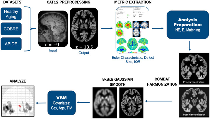
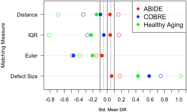
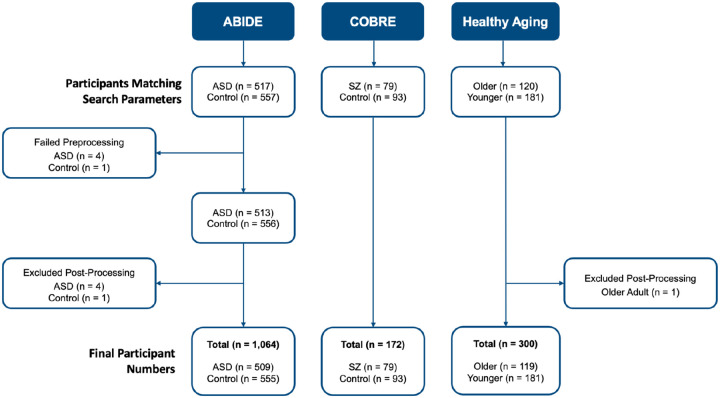
Movement during MRI scanning complicates distinguishing between the different tissues in the brain (e.g., grey and white matter). Standard practice excludes scans based on researcher-determined visual quality thresholds. Unfortunately, children, elderly, and clinical populations are shown to move more, resulting in higher exclusion rates. This disproportionate exclusion creates systematic bias in the literature and makes research findings less generalizable. Furthermore, the artifacts caused by motion are demonstrated to continue to confound data, even after visual quality control has occurred. We aimed to minimize the confounding factor of systematic group differences in movement. To achieve this, we used a post-scanning statistical technique called propensity score matching (PSM) that matches control and patient populations on scan quality metrics, leading to more comparable groups, greater inclusion, and more generalizable results. We found that PSM can attenuate significant differences in scan quality between groups while allowing for greater sample diversity than standard exclusion protocols. Crucially, using PSM can also alter the results of neuroimaging-based analyses. Using three datasets (total n = 1536), we compared voxel based morphometry analyses based on different quality control protocols. In particular, we observed discrepant results between PSM and strict threshold exclusion, with PSM magnifying some regional group differences and diminishing others. Overall, PSM is a customizable way to mitigate the impact of confounds in neuroimaging research and a powerful method to help distinguish true effects from artifacts.

 求助内容:
求助内容: 应助结果提醒方式:


