Grigory Demyashkin, Dmitrii Atiakshin, Kirill Silakov, Vladimir Shchekin, Maxim Bobrov, Olga Abramova, Matvey Vadyukhin, Tatyana Borovaya, Ekaterina Blinova, Petr Shegay, Andrei Kaprin
{"title":"Phenotypic and Quantitative Characterization of Mast Cells in Cutaneous Melanoma: Correlation with Staging Metrics.","authors":"Grigory Demyashkin, Dmitrii Atiakshin, Kirill Silakov, Vladimir Shchekin, Maxim Bobrov, Olga Abramova, Matvey Vadyukhin, Tatyana Borovaya, Ekaterina Blinova, Petr Shegay, Andrei Kaprin","doi":"10.3390/cimb47090752","DOIUrl":null,"url":null,"abstract":"<p><p><b>Background</b>: Mast cells, key effectors of the innate immune system, are known to participate in various stages of tumor progression, including inflammation, angiogenesis, and extracellular matrix remodeling. Their role in melanoma, particularly in relation to Breslow thickness, pT stage, and AJCC staging, remains unclear. This study aims to quantitatively and phenotypically assess mast cell infiltration in cutaneous melanoma at different stages of progression, focusing on Tryptase- and Chymase-positive subtypes. <b>Methods</b>: This retrospective multicenter study included 124 patients with cutaneous melanoma (AJCC 8th edition, stages IA-IIIC). Histological sections were stained with hematoxylin and eosin, and mast cells were visualized using toluidine blue and immunohistochemistry with anti-Tryptase and anti-Chymase antibodies. Mast cells were counted manually in intratumoral and peritumoral regions by two independent observers. Quantitative data were analyzed using non-parametric tests and presented as median [Q1-Q3]. <b>Results</b>: Histological examination of 124 melanoma samples confirmed typical features of cutaneous melanoma, with nodular melanoma being the most common subtype (68 cases, 54.8%) and the lower extremities identified as the predominant tumor location (47 cases, 37.9%). Toluidine blue staining verified the presence of mast cells in both intratumoral and peritumoral compartments, with the highest density observed in early-stage melanomas. Immunohistochemical analysis identified both Tryptase+ and Chymase+ mast cells. The intratumoral number of Tryptase+ cells declined from 17 [14-19] per HPF at AJCC stage IA to 6 [5-7] per HPF at stage IIIC, while Chymase+ mast cells decreased from 14 [11-16] per HPF to 2 [1-3] per HPF over the same stages. Peritumoral counts also showed a downward trend, although less pronounced. Overall, the most significant reduction was observed in Chymase+ mast cells, suggesting their potential role as markers of melanoma progression. <b>Conclusions</b>: This study highlights the dynamic changes in mast cell populations in cutaneous melanoma, with a pronounced decrease in Chymase<sup>+</sup> mast cells as the tumor progresses. Further research is needed to explore the mechanistic role of mast cells and their phenotypic shifts in melanoma progression.</p>","PeriodicalId":10839,"journal":{"name":"Current Issues in Molecular Biology","volume":"47 9","pages":""},"PeriodicalIF":3.0000,"publicationDate":"2025-09-12","publicationTypes":"Journal Article","fieldsOfStudy":null,"isOpenAccess":false,"openAccessPdf":"https://www.ncbi.nlm.nih.gov/pmc/articles/PMC12468969/pdf/","citationCount":"0","resultStr":null,"platform":"Semanticscholar","paperid":null,"PeriodicalName":"Current Issues in Molecular Biology","FirstCategoryId":"99","ListUrlMain":"https://doi.org/10.3390/cimb47090752","RegionNum":3,"RegionCategory":"生物学","ArticlePicture":[],"TitleCN":null,"AbstractTextCN":null,"PMCID":null,"EPubDate":"","PubModel":"","JCR":"Q3","JCRName":"BIOCHEMISTRY & MOLECULAR BIOLOGY","Score":null,"Total":0}
引用次数: 0
Abstract
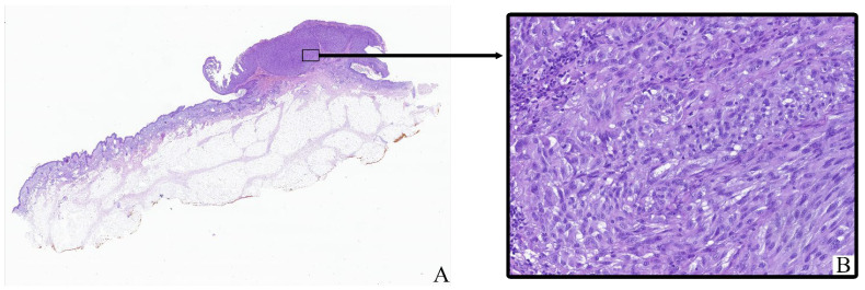
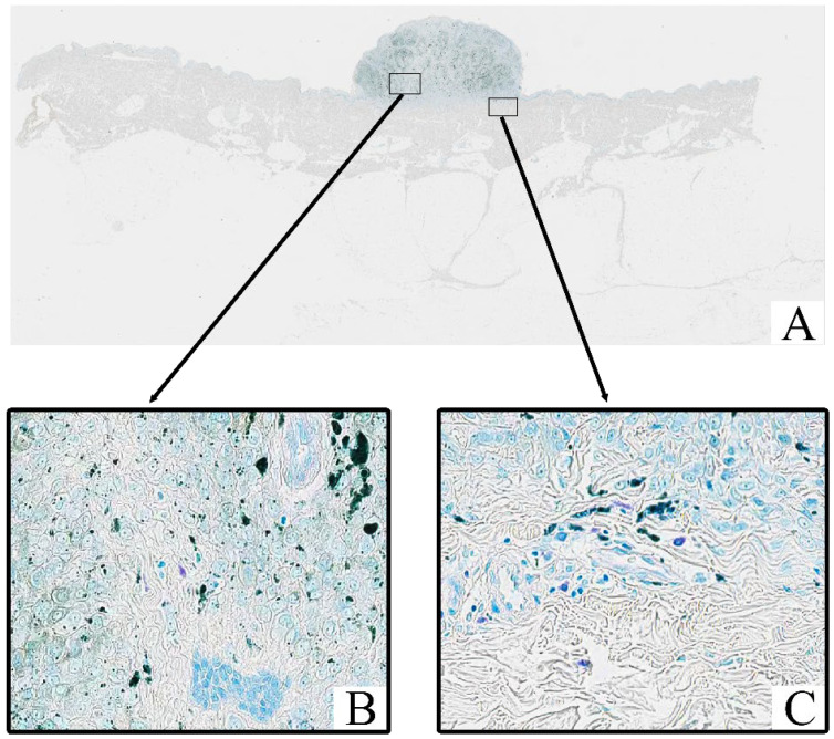
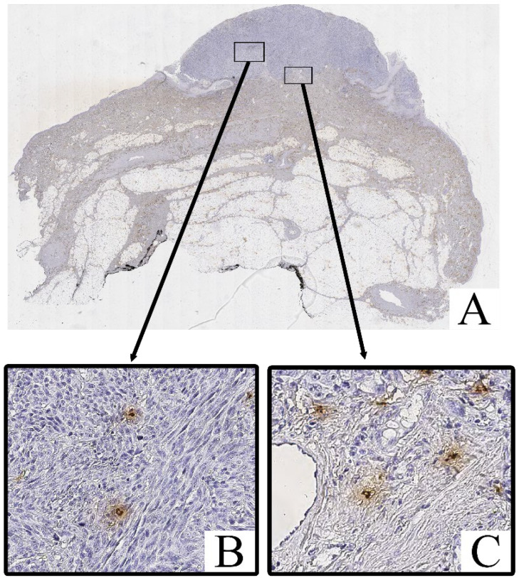
Background: Mast cells, key effectors of the innate immune system, are known to participate in various stages of tumor progression, including inflammation, angiogenesis, and extracellular matrix remodeling. Their role in melanoma, particularly in relation to Breslow thickness, pT stage, and AJCC staging, remains unclear. This study aims to quantitatively and phenotypically assess mast cell infiltration in cutaneous melanoma at different stages of progression, focusing on Tryptase- and Chymase-positive subtypes. Methods: This retrospective multicenter study included 124 patients with cutaneous melanoma (AJCC 8th edition, stages IA-IIIC). Histological sections were stained with hematoxylin and eosin, and mast cells were visualized using toluidine blue and immunohistochemistry with anti-Tryptase and anti-Chymase antibodies. Mast cells were counted manually in intratumoral and peritumoral regions by two independent observers. Quantitative data were analyzed using non-parametric tests and presented as median [Q1-Q3]. Results: Histological examination of 124 melanoma samples confirmed typical features of cutaneous melanoma, with nodular melanoma being the most common subtype (68 cases, 54.8%) and the lower extremities identified as the predominant tumor location (47 cases, 37.9%). Toluidine blue staining verified the presence of mast cells in both intratumoral and peritumoral compartments, with the highest density observed in early-stage melanomas. Immunohistochemical analysis identified both Tryptase+ and Chymase+ mast cells. The intratumoral number of Tryptase+ cells declined from 17 [14-19] per HPF at AJCC stage IA to 6 [5-7] per HPF at stage IIIC, while Chymase+ mast cells decreased from 14 [11-16] per HPF to 2 [1-3] per HPF over the same stages. Peritumoral counts also showed a downward trend, although less pronounced. Overall, the most significant reduction was observed in Chymase+ mast cells, suggesting their potential role as markers of melanoma progression. Conclusions: This study highlights the dynamic changes in mast cell populations in cutaneous melanoma, with a pronounced decrease in Chymase+ mast cells as the tumor progresses. Further research is needed to explore the mechanistic role of mast cells and their phenotypic shifts in melanoma progression.
期刊介绍:
Current Issues in Molecular Biology (CIMB) is a peer-reviewed journal publishing review articles and minireviews in all areas of molecular biology and microbiology. Submitted articles are subject to an Article Processing Charge (APC) and are open access immediately upon publication. All manuscripts undergo a peer-review process.
 求助内容:
求助内容: 应助结果提醒方式:


