Ahmad Pour-Rashidi, Sara Zandpazandi, Laetitia Perronne, Virginia B Hill, Chase Krumpelman, Kamal Subedi, Linda Kelahan, Amir A Borhani, Hatice Savas, Ryan Avery, Tugce Agirlar Trabzonlu, Ulas Bagci, Sean Sachdev, Karan Dixit, Rimas V Lukas, Priya Kumthekar, Yuri S Velichko
{"title":"Intracranial Metastases from Uterine Leiomyosarcoma: A Systematic Review and Case Illustration.","authors":"Ahmad Pour-Rashidi, Sara Zandpazandi, Laetitia Perronne, Virginia B Hill, Chase Krumpelman, Kamal Subedi, Linda Kelahan, Amir A Borhani, Hatice Savas, Ryan Avery, Tugce Agirlar Trabzonlu, Ulas Bagci, Sean Sachdev, Karan Dixit, Rimas V Lukas, Priya Kumthekar, Yuri S Velichko","doi":"10.3390/jcm14186631","DOIUrl":null,"url":null,"abstract":"<p><p><b>Background/Objectives</b>: Brain metastasis from uterine leiomyosarcoma (ULMS) is an exceptionally rare complication of an aggressive malignancy. With fewer than 40 cases previously documented, a significant knowledge gap exists regarding its clinical course, management, and outcomes. This study provides the largest analysis of ULMS brain metastases to date, integrating a systematic literature review with a novel case report illustrating the disease's uniquely rapid progression. <b>Methods</b>: Following PRISMA guidelines, we systematically reviewed four major databases to identify all reported cases of intracranial metastasis from ULMS. Data on patient demographics, clinico-radiological features, treatments, and survival were extracted and analyzed. Methodological quality was assessed using a modified Joanna Briggs Institute (JBI) tool. <b>Results</b>: We analyzed 34 studies with 39 individual cases. Additionally, this review was supplemented by one new illustrative case from our institution. The median patient age was 51.5 years, and most presented with focal neurological symptoms. Common imaging findings included hyperdense lesions on CT and homogeneously enhancing, dural-based masses on MRI, which mimic other intracranial pathologies. Though surgery was the most frequent intervention (76.9%), median survival after a brain metastasis diagnosis was a grim 5 months, with no significant difference observed between treatment modalities. Our illustrative case was remarkable for an extremely rapid volumetric doubling time averaging just 7.3 days. <b>Conclusions</b>: Brain metastasis from ULMS is a lethal event with an extremely poor prognosis. Nonspecific imaging features create diagnostic challenges, necessitating histopathological confirmation. Current therapies, including surgery and radiotherapy, offer palliative benefit but do not significantly alter survival. The aggressive biological behavior demonstrated here underscores the urgent need for increased clinical awareness and collaborative research to develop more effective management strategies and improve outcomes for this devastating diagnosis.</p>","PeriodicalId":15533,"journal":{"name":"Journal of Clinical Medicine","volume":"14 18","pages":""},"PeriodicalIF":2.9000,"publicationDate":"2025-09-20","publicationTypes":"Journal Article","fieldsOfStudy":null,"isOpenAccess":false,"openAccessPdf":"https://www.ncbi.nlm.nih.gov/pmc/articles/PMC12470581/pdf/","citationCount":"0","resultStr":null,"platform":"Semanticscholar","paperid":null,"PeriodicalName":"Journal of Clinical Medicine","FirstCategoryId":"3","ListUrlMain":"https://doi.org/10.3390/jcm14186631","RegionNum":3,"RegionCategory":"医学","ArticlePicture":[],"TitleCN":null,"AbstractTextCN":null,"PMCID":null,"EPubDate":"","PubModel":"","JCR":"Q1","JCRName":"MEDICINE, GENERAL & INTERNAL","Score":null,"Total":0}
引用次数: 0
Abstract
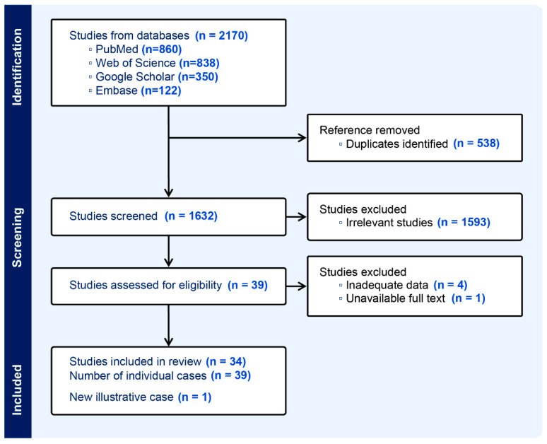
Background/Objectives: Brain metastasis from uterine leiomyosarcoma (ULMS) is an exceptionally rare complication of an aggressive malignancy. With fewer than 40 cases previously documented, a significant knowledge gap exists regarding its clinical course, management, and outcomes. This study provides the largest analysis of ULMS brain metastases to date, integrating a systematic literature review with a novel case report illustrating the disease's uniquely rapid progression. Methods: Following PRISMA guidelines, we systematically reviewed four major databases to identify all reported cases of intracranial metastasis from ULMS. Data on patient demographics, clinico-radiological features, treatments, and survival were extracted and analyzed. Methodological quality was assessed using a modified Joanna Briggs Institute (JBI) tool. Results: We analyzed 34 studies with 39 individual cases. Additionally, this review was supplemented by one new illustrative case from our institution. The median patient age was 51.5 years, and most presented with focal neurological symptoms. Common imaging findings included hyperdense lesions on CT and homogeneously enhancing, dural-based masses on MRI, which mimic other intracranial pathologies. Though surgery was the most frequent intervention (76.9%), median survival after a brain metastasis diagnosis was a grim 5 months, with no significant difference observed between treatment modalities. Our illustrative case was remarkable for an extremely rapid volumetric doubling time averaging just 7.3 days. Conclusions: Brain metastasis from ULMS is a lethal event with an extremely poor prognosis. Nonspecific imaging features create diagnostic challenges, necessitating histopathological confirmation. Current therapies, including surgery and radiotherapy, offer palliative benefit but do not significantly alter survival. The aggressive biological behavior demonstrated here underscores the urgent need for increased clinical awareness and collaborative research to develop more effective management strategies and improve outcomes for this devastating diagnosis.
期刊介绍:
Journal of Clinical Medicine (ISSN 2077-0383), is an international scientific open access journal, providing a platform for advances in health care/clinical practices, the study of direct observation of patients and general medical research. This multi-disciplinary journal is aimed at a wide audience of medical researchers and healthcare professionals.
Unique features of this journal:
manuscripts regarding original research and ideas will be particularly welcomed.JCM also accepts reviews, communications, and short notes.
There is no limit to publication length: our aim is to encourage scientists to publish their experimental and theoretical results in as much detail as possible.
 求助内容:
求助内容: 应助结果提醒方式:


