Stella Marinelli Pedrini, Thiago P A Aloia, André H Aguillera, Paula M P S Gomes, Jamil R Cade, Francisco Sandro Menezes-Rodrigues, Bárbara P Freitas, Marco T Souza, Francisco A H Fonseca, Marcos Danillo Oliveira, Breno O Almeida, Andrey J Serra, Renato D Lopes, Rita Sinigaglia-Coimbra, Adriano Caixeta
{"title":"Optical and Scanning Electron Microscopy Thrombus Findings in Patients with STEMI Undergoing Primary Versus Rescue PCI.","authors":"Stella Marinelli Pedrini, Thiago P A Aloia, André H Aguillera, Paula M P S Gomes, Jamil R Cade, Francisco Sandro Menezes-Rodrigues, Bárbara P Freitas, Marco T Souza, Francisco A H Fonseca, Marcos Danillo Oliveira, Breno O Almeida, Andrey J Serra, Renato D Lopes, Rita Sinigaglia-Coimbra, Adriano Caixeta","doi":"10.3390/biomedicines13092235","DOIUrl":null,"url":null,"abstract":"<p><p><b>Background</b>: The mechanisms underlying fibrinolysis failure in patients with STEMI who are undergoing a pharmacoinvasive strategy appear to be multifactorial and may be associated with the thrombus's architecture and composition. <b>Objective</b>: We aimed to compare the thrombus composition in patients with STEMI who were undergoing rescue percutaneous coronary intervention (rPCI) versus primary PCI (pPCI) using optical microscopy (OM) and scanning electron microscopy (SEM). Methods: Fifty-three patients were prospectively enrolled, with twenty-five undergoing rPCI and twenty-eight undergoing pPCI. After thrombus aspiration, each harvested fragment was divided into two pieces: one was analyzed using OM with a 60× magnifying lens on hematoxylin-eosin-stained samples, and the other with SEM at 5000× magnification. <b>Results</b>: Patients who underwent rPCI had significantly higher C-reactive protein levels and a longer ischemic interval at admission compared to those treated with pPCI (9.92 h [range: 1.58-106.17] vs. 2.14 h [range: 0-48]; <i>p</i> < 0.001). Optical microscopy analysis revealed that thrombi from rPCI patients exhibited a significantly higher erythrocyte area percentage (18.36% [range: 0.3-50.08] vs. 0.91% [range: 0-70.1]; <i>p</i> = 0.001), a lower fibrin content as assessed by optical microscopy (79.49% [range: 49.2-98.25] vs. 94.43% [range: 29.19-99.92]; <i>p</i> = 0.006), and a greater amount of cholesterol crystals as measured by SEM (1.73 μm<sup>2</sup> [range: 0-18.51] vs. 0.08 μm<sup>2</sup> [range: 0-0.71]; <i>p</i> < 0.001). <b>Conclusions</b>: The thrombus composition of patients with STEMI who are undergoing rPCI had higher amounts of erythrocytes and cholesterol crystals and a lesser area occupied by fibrin compared to those undergoing pPCI. The composition of thrombi in rPCI could potentially contribute to the failure of fibrinolytic therapy within a pharmacoinvasive strategy.</p>","PeriodicalId":8937,"journal":{"name":"Biomedicines","volume":"13 9","pages":""},"PeriodicalIF":3.9000,"publicationDate":"2025-09-11","publicationTypes":"Journal Article","fieldsOfStudy":null,"isOpenAccess":false,"openAccessPdf":"https://www.ncbi.nlm.nih.gov/pmc/articles/PMC12467207/pdf/","citationCount":"0","resultStr":null,"platform":"Semanticscholar","paperid":null,"PeriodicalName":"Biomedicines","FirstCategoryId":"5","ListUrlMain":"https://doi.org/10.3390/biomedicines13092235","RegionNum":3,"RegionCategory":"工程技术","ArticlePicture":[],"TitleCN":null,"AbstractTextCN":null,"PMCID":null,"EPubDate":"","PubModel":"","JCR":"Q2","JCRName":"BIOCHEMISTRY & MOLECULAR BIOLOGY","Score":null,"Total":0}
引用次数: 0
Abstract
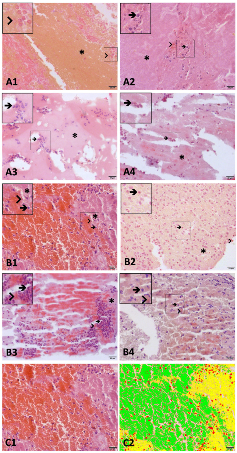
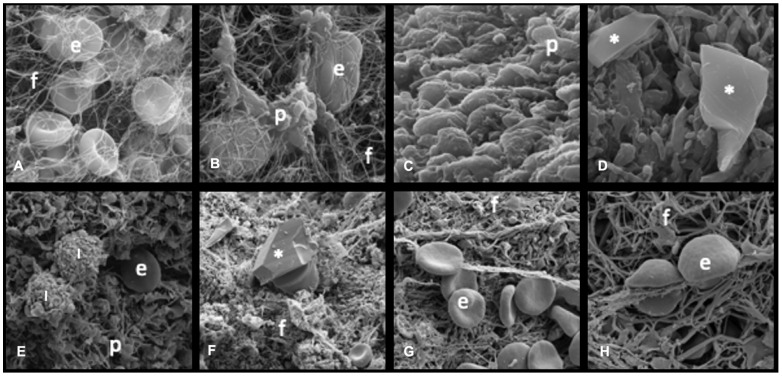
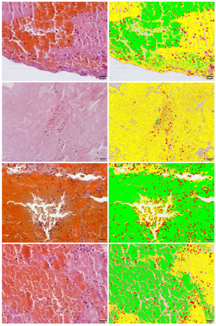
Background: The mechanisms underlying fibrinolysis failure in patients with STEMI who are undergoing a pharmacoinvasive strategy appear to be multifactorial and may be associated with the thrombus's architecture and composition. Objective: We aimed to compare the thrombus composition in patients with STEMI who were undergoing rescue percutaneous coronary intervention (rPCI) versus primary PCI (pPCI) using optical microscopy (OM) and scanning electron microscopy (SEM). Methods: Fifty-three patients were prospectively enrolled, with twenty-five undergoing rPCI and twenty-eight undergoing pPCI. After thrombus aspiration, each harvested fragment was divided into two pieces: one was analyzed using OM with a 60× magnifying lens on hematoxylin-eosin-stained samples, and the other with SEM at 5000× magnification. Results: Patients who underwent rPCI had significantly higher C-reactive protein levels and a longer ischemic interval at admission compared to those treated with pPCI (9.92 h [range: 1.58-106.17] vs. 2.14 h [range: 0-48]; p < 0.001). Optical microscopy analysis revealed that thrombi from rPCI patients exhibited a significantly higher erythrocyte area percentage (18.36% [range: 0.3-50.08] vs. 0.91% [range: 0-70.1]; p = 0.001), a lower fibrin content as assessed by optical microscopy (79.49% [range: 49.2-98.25] vs. 94.43% [range: 29.19-99.92]; p = 0.006), and a greater amount of cholesterol crystals as measured by SEM (1.73 μm2 [range: 0-18.51] vs. 0.08 μm2 [range: 0-0.71]; p < 0.001). Conclusions: The thrombus composition of patients with STEMI who are undergoing rPCI had higher amounts of erythrocytes and cholesterol crystals and a lesser area occupied by fibrin compared to those undergoing pPCI. The composition of thrombi in rPCI could potentially contribute to the failure of fibrinolytic therapy within a pharmacoinvasive strategy.
BiomedicinesBiochemistry, Genetics and Molecular Biology-General Biochemistry,Genetics and Molecular Biology
CiteScore
5.20
自引率
8.50%
发文量
2823
审稿时长
8 weeks
期刊介绍:
Biomedicines (ISSN 2227-9059; CODEN: BIOMID) is an international, scientific, open access journal on biomedicines published quarterly online by MDPI.
 求助内容:
求助内容: 应助结果提醒方式:


