{"title":"Structural Proteins at Neuromuscular Junction Are Downgraded While NRG1 and Agrin Gene Expression Increases After Muscle Injury.","authors":"Jurandyr Pimentel Neto, Lara Caetano Rocha-Braga, Matheus Bertanha Fior, Paula Oliveira Camargo, Adriano Polican Ciena","doi":"10.3390/biomedicines13092277","DOIUrl":null,"url":null,"abstract":"<p><p><b>Background/Objectives:</b> The neuromuscular junction (NMJ) is the area where peripheral nerves communicate with muscle tissue. Muscle injury can occur as part of an acute degenerative process at the NMJ. This study aims to investigate the remodeling of the NMJ after a muscle injury in an experimental model. <b>Methods:</b> We used sixty male Wistar rats divided into five groups: a control group (C) and four muscle injury groups (MI) at different time points: 0 h, 24 h, 48 h, and 7 d after injury. We subjected the right hind limb to muscle injury and dissected the gastrocnemius muscles for analysis. We employed light microscopy to examine cell nuclei and the connective tissue, immunostaining to identify and measure the pre- and postsynaptic regions as well as calcium channels (P/Q), and real-time PCR to assess the gene expression of NRG1 and Agrin. <b>Results:</b> Our findings revealed an accumulation of nuclei and connective tissue in the acute injury groups (0 to 48 h). The morpho-quantitative analyses showed that the presynaptic structures and calcium channels underwent similar remodeling due to their morpho-functional relationship. Meanwhile, the postsynaptic receptors were significantly affected by the degenerative and inflammatory processes. These results can be linked to increased expression of NRG1 and Agrin in the acute injury groups. <b>Conclusions:</b> In conclusion, the synaptic regions displayed substantial adaptations within the first 48 h, with the presynaptic region recovering rapidly and the postsynaptic region recovering slowly. This relationship suggests that significant increases in Agrin and NRG1 play a crucial role in maintaining the integrity of these structures.</p>","PeriodicalId":8937,"journal":{"name":"Biomedicines","volume":"13 9","pages":""},"PeriodicalIF":3.9000,"publicationDate":"2025-09-16","publicationTypes":"Journal Article","fieldsOfStudy":null,"isOpenAccess":false,"openAccessPdf":"https://www.ncbi.nlm.nih.gov/pmc/articles/PMC12467557/pdf/","citationCount":"0","resultStr":null,"platform":"Semanticscholar","paperid":null,"PeriodicalName":"Biomedicines","FirstCategoryId":"5","ListUrlMain":"https://doi.org/10.3390/biomedicines13092277","RegionNum":3,"RegionCategory":"工程技术","ArticlePicture":[],"TitleCN":null,"AbstractTextCN":null,"PMCID":null,"EPubDate":"","PubModel":"","JCR":"Q2","JCRName":"BIOCHEMISTRY & MOLECULAR BIOLOGY","Score":null,"Total":0}
引用次数: 0
Abstract
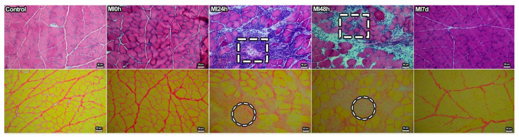
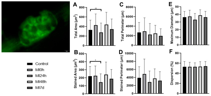
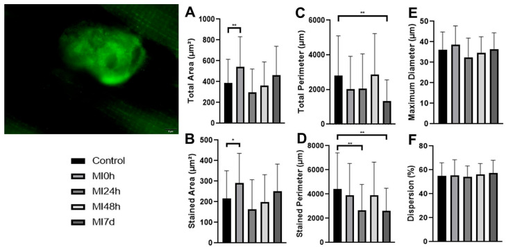
Background/Objectives: The neuromuscular junction (NMJ) is the area where peripheral nerves communicate with muscle tissue. Muscle injury can occur as part of an acute degenerative process at the NMJ. This study aims to investigate the remodeling of the NMJ after a muscle injury in an experimental model. Methods: We used sixty male Wistar rats divided into five groups: a control group (C) and four muscle injury groups (MI) at different time points: 0 h, 24 h, 48 h, and 7 d after injury. We subjected the right hind limb to muscle injury and dissected the gastrocnemius muscles for analysis. We employed light microscopy to examine cell nuclei and the connective tissue, immunostaining to identify and measure the pre- and postsynaptic regions as well as calcium channels (P/Q), and real-time PCR to assess the gene expression of NRG1 and Agrin. Results: Our findings revealed an accumulation of nuclei and connective tissue in the acute injury groups (0 to 48 h). The morpho-quantitative analyses showed that the presynaptic structures and calcium channels underwent similar remodeling due to their morpho-functional relationship. Meanwhile, the postsynaptic receptors were significantly affected by the degenerative and inflammatory processes. These results can be linked to increased expression of NRG1 and Agrin in the acute injury groups. Conclusions: In conclusion, the synaptic regions displayed substantial adaptations within the first 48 h, with the presynaptic region recovering rapidly and the postsynaptic region recovering slowly. This relationship suggests that significant increases in Agrin and NRG1 play a crucial role in maintaining the integrity of these structures.
BiomedicinesBiochemistry, Genetics and Molecular Biology-General Biochemistry,Genetics and Molecular Biology
CiteScore
5.20
自引率
8.50%
发文量
2823
审稿时长
8 weeks
期刊介绍:
Biomedicines (ISSN 2227-9059; CODEN: BIOMID) is an international, scientific, open access journal on biomedicines published quarterly online by MDPI.
 求助内容:
求助内容: 应助结果提醒方式:


