V. V. Martynova, S. V. Akulinichev, I. A. Yakovlev
{"title":"Analysis of Cell Death and Proliferative Activity of Cell Cultures under Proton Irradiation in Flash Mode","authors":"V. V. Martynova, S. V. Akulinichev, I. A. Yakovlev","doi":"10.3103/S0027134925700353","DOIUrl":null,"url":null,"abstract":"<p>The study of the biological effects of accelerated protons with ultrahigh dose rates in and outside the Bragg peak on tumour and normal cell lines is essential for understanding the consequences of such exposure and for selecting optimal doses and modes for further application in tumour radiotherapy. In this study, human colorectal adenocarcinoma cells (HT-29) and normal human adipose-derived mesenchymal stem cells (ADSC)—fibroblasts—were irradiated. The irradiation was performed using the high-current linear proton accelerator at the Institute for Nuclear Research of the Russian Academy of Sciences, which allows for energy variation in the range of 70–230 MeV. The dose was delivered in three modes: conventional mode (dose rate <span>\\(\\dot{D}<1\\)</span> Gy/s), Splash mode (<span>\\(\\dot{D}\\sim 100\\)</span> Gy/s), and single-pulse Flash mode (<span>\\(\\dot{D}>10^{4}\\)</span> Gy/s, denoted as Splash from single-pulse Flash) in the region of the spread out Bragg peak. To analyze cell death, staining with propidium iodide and annexin was performed. The proliferative potential was assessed using the EdU assay. After 24 h, an increase in the number of apoptotic HT-29 cells was observed in all irradiation modes, while for fibroblasts, a relative increase in the number of necrotic cells was noted under conventional irradiation with a higher dose compared to other modes. After 48 h, a tendency toward a dose-dependent decrease in the number of necrotic HT-29 cells was observed in Flash/Splash modes, with consistently low levels of necrotic fibroblasts. The number of DNA-synthesizing cells decreased significantly by 120 h across all doses and irradiation modes. The curves for Flash and Splash irradiation modes were nearly parallel. Further studies involving a broader range of cell lines and doses are required.</p>","PeriodicalId":711,"journal":{"name":"Moscow University Physics Bulletin","volume":"80 2","pages":"270 - 276"},"PeriodicalIF":0.4000,"publicationDate":"2025-07-13","publicationTypes":"Journal Article","fieldsOfStudy":null,"isOpenAccess":false,"openAccessPdf":"","citationCount":"0","resultStr":null,"platform":"Semanticscholar","paperid":null,"PeriodicalName":"Moscow University Physics Bulletin","FirstCategoryId":"101","ListUrlMain":"https://link.springer.com/article/10.3103/S0027134925700353","RegionNum":4,"RegionCategory":"物理与天体物理","ArticlePicture":[],"TitleCN":null,"AbstractTextCN":null,"PMCID":null,"EPubDate":"","PubModel":"","JCR":"Q4","JCRName":"PHYSICS, MULTIDISCIPLINARY","Score":null,"Total":0}
引用次数: 0
Abstract
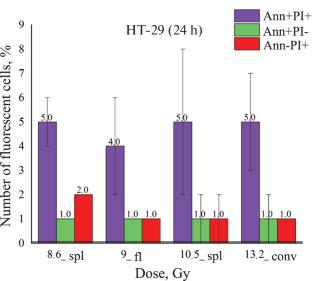
The study of the biological effects of accelerated protons with ultrahigh dose rates in and outside the Bragg peak on tumour and normal cell lines is essential for understanding the consequences of such exposure and for selecting optimal doses and modes for further application in tumour radiotherapy. In this study, human colorectal adenocarcinoma cells (HT-29) and normal human adipose-derived mesenchymal stem cells (ADSC)—fibroblasts—were irradiated. The irradiation was performed using the high-current linear proton accelerator at the Institute for Nuclear Research of the Russian Academy of Sciences, which allows for energy variation in the range of 70–230 MeV. The dose was delivered in three modes: conventional mode (dose rate \(\dot{D}<1\) Gy/s), Splash mode (\(\dot{D}\sim 100\) Gy/s), and single-pulse Flash mode (\(\dot{D}>10^{4}\) Gy/s, denoted as Splash from single-pulse Flash) in the region of the spread out Bragg peak. To analyze cell death, staining with propidium iodide and annexin was performed. The proliferative potential was assessed using the EdU assay. After 24 h, an increase in the number of apoptotic HT-29 cells was observed in all irradiation modes, while for fibroblasts, a relative increase in the number of necrotic cells was noted under conventional irradiation with a higher dose compared to other modes. After 48 h, a tendency toward a dose-dependent decrease in the number of necrotic HT-29 cells was observed in Flash/Splash modes, with consistently low levels of necrotic fibroblasts. The number of DNA-synthesizing cells decreased significantly by 120 h across all doses and irradiation modes. The curves for Flash and Splash irradiation modes were nearly parallel. Further studies involving a broader range of cell lines and doses are required.
期刊介绍:
Moscow University Physics Bulletin publishes original papers (reviews, articles, and brief communications) in the following fields of experimental and theoretical physics: theoretical and mathematical physics; physics of nuclei and elementary particles; radiophysics, electronics, acoustics; optics and spectroscopy; laser physics; condensed matter physics; chemical physics, physical kinetics, and plasma physics; biophysics and medical physics; astronomy, astrophysics, and cosmology; physics of the Earth’s, atmosphere, and hydrosphere.
 求助内容:
求助内容: 应助结果提醒方式:


