{"title":"Minimally Invasive Treatment of Traumatic Diaphragm Rupture.","authors":"Mustafa Yılmaz, Cansu Zerey, Hilmi Bozkurt","doi":"10.4293/CRSLS.2025.00071","DOIUrl":null,"url":null,"abstract":"<p><strong>Background: </strong>Diaphragmatic rupture is a rare but potentially life-threatening injury that can result from blunt or penetrating trauma. Timely diagnosis is critical to prevent serious complications, yet clinical and radiological assessments are often inconclusive.</p><p><strong>Case presentation: </strong>We report the case of a 39-year-old male patient who sustained blunt thoracoabdominal trauma in a motor vehicle accident. The patient presented with respiratory distress and generalized abdominal tenderness. Imaging revealed a left diaphragmatic defect with herniation of abdominal organs into the thoracic cavity.</p><p><strong>Surgical technique: </strong>The patient underwent diagnostic laparoscopy, which confirmed an 8 × 5 cm defect in the left diaphragmatic dome with herniation of the stomach and omentum. The herniated organs were reduced, and the defect was repaired tension-free using intracorporeal 0-silk sutures. A 28-Fr intercostal drainage tube and a 12-Fr abdominal drain were placed.</p><p><strong>Outcome: </strong>The postoperative course was uneventful. A chest x-ray on postoperative day one confirmed normal diaphragm position and re-expansion of the lung. The patient recovered without complications.</p><p><strong>Conclusion: </strong>Laparoscopy provides a minimally invasive and effective diagnostic and therapeutic option for diaphragmatic rupture following blunt trauma. In appropriately selected cases, it offers favorable outcomes with reduced morbidity.</p>","PeriodicalId":72723,"journal":{"name":"CRSLS : MIS case reports from SLS","volume":"12 3","pages":""},"PeriodicalIF":0.0000,"publicationDate":"2025-09-24","publicationTypes":"Journal Article","fieldsOfStudy":null,"isOpenAccess":false,"openAccessPdf":"https://www.ncbi.nlm.nih.gov/pmc/articles/PMC12458920/pdf/","citationCount":"0","resultStr":null,"platform":"Semanticscholar","paperid":null,"PeriodicalName":"CRSLS : MIS case reports from SLS","FirstCategoryId":"1085","ListUrlMain":"https://doi.org/10.4293/CRSLS.2025.00071","RegionNum":0,"RegionCategory":null,"ArticlePicture":[],"TitleCN":null,"AbstractTextCN":null,"PMCID":null,"EPubDate":"2025/7/1 0:00:00","PubModel":"eCollection","JCR":"","JCRName":"","Score":null,"Total":0}
引用次数: 0
Abstract
Background: Diaphragmatic rupture is a rare but potentially life-threatening injury that can result from blunt or penetrating trauma. Timely diagnosis is critical to prevent serious complications, yet clinical and radiological assessments are often inconclusive.
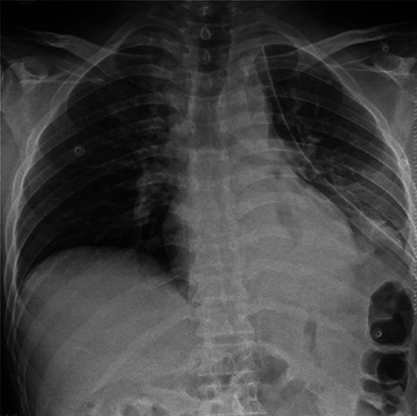
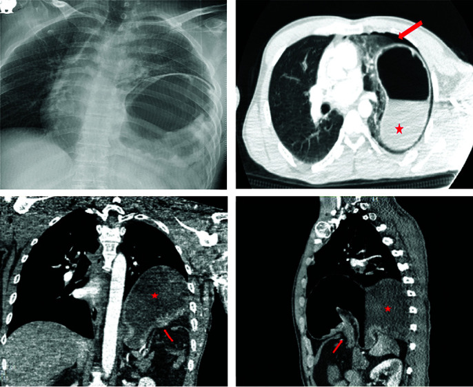
Case presentation: We report the case of a 39-year-old male patient who sustained blunt thoracoabdominal trauma in a motor vehicle accident. The patient presented with respiratory distress and generalized abdominal tenderness. Imaging revealed a left diaphragmatic defect with herniation of abdominal organs into the thoracic cavity.
Surgical technique: The patient underwent diagnostic laparoscopy, which confirmed an 8 × 5 cm defect in the left diaphragmatic dome with herniation of the stomach and omentum. The herniated organs were reduced, and the defect was repaired tension-free using intracorporeal 0-silk sutures. A 28-Fr intercostal drainage tube and a 12-Fr abdominal drain were placed.
Outcome: The postoperative course was uneventful. A chest x-ray on postoperative day one confirmed normal diaphragm position and re-expansion of the lung. The patient recovered without complications.
Conclusion: Laparoscopy provides a minimally invasive and effective diagnostic and therapeutic option for diaphragmatic rupture following blunt trauma. In appropriately selected cases, it offers favorable outcomes with reduced morbidity.


 求助内容:
求助内容: 应助结果提醒方式:


