Yashbir Singh, Jesper B Andersen, Quincy A Hathaway, Diana V Vera-Garcia, Varekan Keishing, Sudhakar K Venkatesh, Sara Salehi, Davide Povero, Michael B Wallace, Gregory J Gores, Yujia Wei, Natally Horvat, Bradley J Erickson, Emilio Quaia
{"title":"Leveraging Multimodal Foundation Models in Biliary Tract Cancer Research.","authors":"Yashbir Singh, Jesper B Andersen, Quincy A Hathaway, Diana V Vera-Garcia, Varekan Keishing, Sudhakar K Venkatesh, Sara Salehi, Davide Povero, Michael B Wallace, Gregory J Gores, Yujia Wei, Natally Horvat, Bradley J Erickson, Emilio Quaia","doi":"10.3390/tomography11090096","DOIUrl":null,"url":null,"abstract":"<p><p>This review explores how multimodal foundation models (MFMs) are transforming biliary tract cancer (BTC) research. BTCs are aggressive malignancies with poor prognosis, presenting unique challenges due to difficult diagnostic methods, molecular complexity, and rarity. Importantly, intrahepatic cholangiocarcinoma (iCCA), perihilar cholangiocarcinoma (pCCA), and distal bile duct cholangiocarcinoma (dCCA) represent fundamentally distinct clinical entities, with iCCA presenting as mass-forming lesions amenable to biopsy and targeted therapies, while pCCA manifests as infiltrative bile duct lesions with challenging diagnosis and primarily palliative management approaches. MFMs offer potential to advance research by integrating radiological images, histopathology, multi-omics profiles, and clinical data into unified computational frameworks, with applications tailored to these distinct BTC subtypes. Key applications include enhanced biomarker discovery that identifies previously unrecognizable cross-modal patterns, potential for improving currently limited diagnostic accuracy-though validation in BTC-specific cohorts remains essential-accelerated drug repurposing, and advanced patient stratification for personalized treatment. Despite promising results, challenges such as data scarcity, high computational demands, and clinical workflow integration remain to be addressed. Future research should focus on standardized data protocols, architectural innovations, and prospective validation studies. The integration of artificial intelligence (AI)-based methodologies offers new solutions for these historically challenging malignancies. However, current evidence for BTC-specific applications remains largely theoretical, with most studies limited to proof-of-concept designs or related cancer types. Comprehensive clinical validation studies and prospective trials demonstrating patient benefit are essential prerequisites for clinical implementation. The timeline for evidence-based clinical adoption likely extends 7-10 years, contingent on successful completion of validation studies addressing current evidence gaps.</p>","PeriodicalId":51330,"journal":{"name":"Tomography","volume":"11 9","pages":""},"PeriodicalIF":2.2000,"publicationDate":"2025-08-25","publicationTypes":"Journal Article","fieldsOfStudy":null,"isOpenAccess":false,"openAccessPdf":"https://www.ncbi.nlm.nih.gov/pmc/articles/PMC12473628/pdf/","citationCount":"0","resultStr":null,"platform":"Semanticscholar","paperid":null,"PeriodicalName":"Tomography","FirstCategoryId":"3","ListUrlMain":"https://doi.org/10.3390/tomography11090096","RegionNum":4,"RegionCategory":"医学","ArticlePicture":[],"TitleCN":null,"AbstractTextCN":null,"PMCID":null,"EPubDate":"","PubModel":"","JCR":"Q2","JCRName":"RADIOLOGY, NUCLEAR MEDICINE & MEDICAL IMAGING","Score":null,"Total":0}
引用次数: 0
Abstract
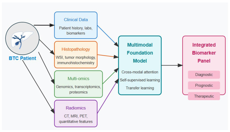
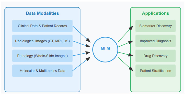
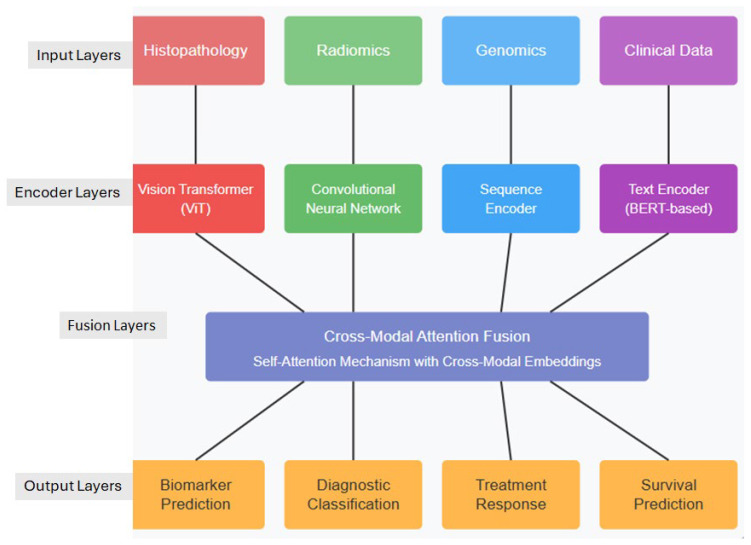
This review explores how multimodal foundation models (MFMs) are transforming biliary tract cancer (BTC) research. BTCs are aggressive malignancies with poor prognosis, presenting unique challenges due to difficult diagnostic methods, molecular complexity, and rarity. Importantly, intrahepatic cholangiocarcinoma (iCCA), perihilar cholangiocarcinoma (pCCA), and distal bile duct cholangiocarcinoma (dCCA) represent fundamentally distinct clinical entities, with iCCA presenting as mass-forming lesions amenable to biopsy and targeted therapies, while pCCA manifests as infiltrative bile duct lesions with challenging diagnosis and primarily palliative management approaches. MFMs offer potential to advance research by integrating radiological images, histopathology, multi-omics profiles, and clinical data into unified computational frameworks, with applications tailored to these distinct BTC subtypes. Key applications include enhanced biomarker discovery that identifies previously unrecognizable cross-modal patterns, potential for improving currently limited diagnostic accuracy-though validation in BTC-specific cohorts remains essential-accelerated drug repurposing, and advanced patient stratification for personalized treatment. Despite promising results, challenges such as data scarcity, high computational demands, and clinical workflow integration remain to be addressed. Future research should focus on standardized data protocols, architectural innovations, and prospective validation studies. The integration of artificial intelligence (AI)-based methodologies offers new solutions for these historically challenging malignancies. However, current evidence for BTC-specific applications remains largely theoretical, with most studies limited to proof-of-concept designs or related cancer types. Comprehensive clinical validation studies and prospective trials demonstrating patient benefit are essential prerequisites for clinical implementation. The timeline for evidence-based clinical adoption likely extends 7-10 years, contingent on successful completion of validation studies addressing current evidence gaps.
TomographyMedicine-Radiology, Nuclear Medicine and Imaging
CiteScore
2.70
自引率
10.50%
发文量
222
期刊介绍:
TomographyTM publishes basic (technical and pre-clinical) and clinical scientific articles which involve the advancement of imaging technologies. Tomography encompasses studies that use single or multiple imaging modalities including for example CT, US, PET, SPECT, MR and hyperpolarization technologies, as well as optical modalities (i.e. bioluminescence, photoacoustic, endomicroscopy, fiber optic imaging and optical computed tomography) in basic sciences, engineering, preclinical and clinical medicine.
Tomography also welcomes studies involving exploration and refinement of contrast mechanisms and image-derived metrics within and across modalities toward the development of novel imaging probes for image-based feedback and intervention. The use of imaging in biology and medicine provides unparalleled opportunities to noninvasively interrogate tissues to obtain real-time dynamic and quantitative information required for diagnosis and response to interventions and to follow evolving pathological conditions. As multi-modal studies and the complexities of imaging technologies themselves are ever increasing to provide advanced information to scientists and clinicians.
Tomography provides a unique publication venue allowing investigators the opportunity to more precisely communicate integrated findings related to the diverse and heterogeneous features associated with underlying anatomical, physiological, functional, metabolic and molecular genetic activities of normal and diseased tissue. Thus Tomography publishes peer-reviewed articles which involve the broad use of imaging of any tissue and disease type including both preclinical and clinical investigations. In addition, hardware/software along with chemical and molecular probe advances are welcome as they are deemed to significantly contribute towards the long-term goal of improving the overall impact of imaging on scientific and clinical discovery.

 求助内容:
求助内容: 应助结果提醒方式:


