{"title":"A Flexible Multi-Channel Deep Network Leveraging Texture and Spatial Features for Diagnosing New COVID-19 Variants in Lung CT Scans.","authors":"Shervan Fekri-Ershad, Khalegh Behrouz Dehkordi","doi":"10.3390/tomography11090099","DOIUrl":null,"url":null,"abstract":"<p><strong>Background: </strong>The COVID-19 pandemic has claimed thousands of lives worldwide. While infection rates have declined in recent years, emerging variants remain a deadly threat. Accurate diagnosis is critical to curbing transmission and improving treatment outcomes. However, the similarity of COVID-19 symptoms to those of the common cold and flu has spurred the development of automated diagnostic methods, particularly through lung computed-tomography (CT) scan analysis.</p><p><strong>Methodology: </strong>This paper proposes a novel deep learning-based approach for detecting diverse COVID-19 variants using advanced textural feature extraction. The framework employs a dual-channel convolutional neural network (CNN), where one channel processes texture-based features and the other analyzes spatial information. Unlike existing methods, our model dynamically learns textural patterns during training, eliminating reliance on predefined features. A modified local binary pattern (LBP) technique extracts texture data in matrix form, while the CNN's adaptable internal architecture optimizes the balance between accuracy and computational efficiency. To enhance performance, hyperparameters are fine-tuned using the Adam optimizer and focal loss function.</p><p><strong>Results: </strong>The proposed method is evaluated on two benchmark datasets, COVID-349 and Italian COVID-Set, which include diverse COVID-19 variants.</p><p><strong>Conclusions: </strong>The results demonstrate its superior accuracy (94.63% and 95.47%, respectively), outperforming competing approaches in precision, recall, and overall diagnostic reliability.</p>","PeriodicalId":51330,"journal":{"name":"Tomography","volume":"11 9","pages":""},"PeriodicalIF":2.2000,"publicationDate":"2025-08-27","publicationTypes":"Journal Article","fieldsOfStudy":null,"isOpenAccess":false,"openAccessPdf":"https://www.ncbi.nlm.nih.gov/pmc/articles/PMC12473366/pdf/","citationCount":"0","resultStr":null,"platform":"Semanticscholar","paperid":null,"PeriodicalName":"Tomography","FirstCategoryId":"3","ListUrlMain":"https://doi.org/10.3390/tomography11090099","RegionNum":4,"RegionCategory":"医学","ArticlePicture":[],"TitleCN":null,"AbstractTextCN":null,"PMCID":null,"EPubDate":"","PubModel":"","JCR":"Q2","JCRName":"RADIOLOGY, NUCLEAR MEDICINE & MEDICAL IMAGING","Score":null,"Total":0}
引用次数: 0
Abstract
Background: The COVID-19 pandemic has claimed thousands of lives worldwide. While infection rates have declined in recent years, emerging variants remain a deadly threat. Accurate diagnosis is critical to curbing transmission and improving treatment outcomes. However, the similarity of COVID-19 symptoms to those of the common cold and flu has spurred the development of automated diagnostic methods, particularly through lung computed-tomography (CT) scan analysis.
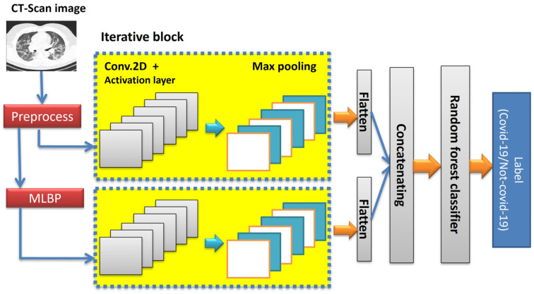
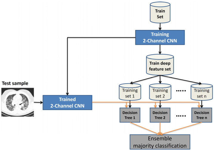
Methodology: This paper proposes a novel deep learning-based approach for detecting diverse COVID-19 variants using advanced textural feature extraction. The framework employs a dual-channel convolutional neural network (CNN), where one channel processes texture-based features and the other analyzes spatial information. Unlike existing methods, our model dynamically learns textural patterns during training, eliminating reliance on predefined features. A modified local binary pattern (LBP) technique extracts texture data in matrix form, while the CNN's adaptable internal architecture optimizes the balance between accuracy and computational efficiency. To enhance performance, hyperparameters are fine-tuned using the Adam optimizer and focal loss function.
Results: The proposed method is evaluated on two benchmark datasets, COVID-349 and Italian COVID-Set, which include diverse COVID-19 variants.
Conclusions: The results demonstrate its superior accuracy (94.63% and 95.47%, respectively), outperforming competing approaches in precision, recall, and overall diagnostic reliability.
TomographyMedicine-Radiology, Nuclear Medicine and Imaging
CiteScore
2.70
自引率
10.50%
发文量
222
期刊介绍:
TomographyTM publishes basic (technical and pre-clinical) and clinical scientific articles which involve the advancement of imaging technologies. Tomography encompasses studies that use single or multiple imaging modalities including for example CT, US, PET, SPECT, MR and hyperpolarization technologies, as well as optical modalities (i.e. bioluminescence, photoacoustic, endomicroscopy, fiber optic imaging and optical computed tomography) in basic sciences, engineering, preclinical and clinical medicine.
Tomography also welcomes studies involving exploration and refinement of contrast mechanisms and image-derived metrics within and across modalities toward the development of novel imaging probes for image-based feedback and intervention. The use of imaging in biology and medicine provides unparalleled opportunities to noninvasively interrogate tissues to obtain real-time dynamic and quantitative information required for diagnosis and response to interventions and to follow evolving pathological conditions. As multi-modal studies and the complexities of imaging technologies themselves are ever increasing to provide advanced information to scientists and clinicians.
Tomography provides a unique publication venue allowing investigators the opportunity to more precisely communicate integrated findings related to the diverse and heterogeneous features associated with underlying anatomical, physiological, functional, metabolic and molecular genetic activities of normal and diseased tissue. Thus Tomography publishes peer-reviewed articles which involve the broad use of imaging of any tissue and disease type including both preclinical and clinical investigations. In addition, hardware/software along with chemical and molecular probe advances are welcome as they are deemed to significantly contribute towards the long-term goal of improving the overall impact of imaging on scientific and clinical discovery.

 求助内容:
求助内容: 应助结果提醒方式:


