Wonju Hong, Jaewoong Kang, So Eui Kim, Taikyeong Jeong, Chang Jin Yoon, In Jae Lee, Lyo Min Kwon, Bum-Joo Cho
{"title":"Deep Learning-Based Diagnosis of Femoropopliteal Artery Steno-Occlusion Using Maximum Intensity Projection Images of CT Angiography.","authors":"Wonju Hong, Jaewoong Kang, So Eui Kim, Taikyeong Jeong, Chang Jin Yoon, In Jae Lee, Lyo Min Kwon, Bum-Joo Cho","doi":"10.3390/tomography11090104","DOIUrl":null,"url":null,"abstract":"<p><p><b>Background/Objectives</b>: To develop and validate deep learning-based models for detecting significant steno-occlusion (SSO)-defined as luminal narrowing greater than 50%-of the femoropopliteal arteries using maximum intensity projection (MIP) images from lower extremity CT angiography (CTA). <b>Methods</b>: This retrospective study utilized MIP images of lower extremity CTA performed between January 2021 and December 2023 for internal model development. Deep learning-based models were developed sequentially to diagnose SSO: screening with single anteroposterior image, followed by four-segment rotational analysis that divided each femoropopliteal artery into four segments and incorporated multi-angle images. Given the cropped images and the shape of stenosis, models were trained to classify the presence of SSO. A temporal validation dataset comprised MIP images from lower extremity CTA performed between January and June 2024. <b>Results</b>: In total, 56,496 segment images from 642 patients (mean age: 68.2 ± 13.5 years; 472 men) were included in the internal dataset. In the single-image analysis, RDNet achieved the highest mean AUC of 0.886 for SSO detection. In the four-segment rotational analysis, RDNet also demonstrated the highest mean AUC, reaching 0.964 in both half-set and full-set approaches. While RDNet recorded the highest mean AUC, all other models showed improved AUCs as the number of input images increased (<i>p</i> < 0.05). In the temporal validation dataset, RDNet again achieved the highest mean AUC (0.959) in the half-set analysis. <b>Conclusions</b>: The deep learning-based model, particularly RDNet, demonstrated excellent performance in detecting SSO of peripheral arteries on MIP images from lower extremity CTA.</p>","PeriodicalId":51330,"journal":{"name":"Tomography","volume":"11 9","pages":""},"PeriodicalIF":2.2000,"publicationDate":"2025-09-08","publicationTypes":"Journal Article","fieldsOfStudy":null,"isOpenAccess":false,"openAccessPdf":"https://www.ncbi.nlm.nih.gov/pmc/articles/PMC12473302/pdf/","citationCount":"0","resultStr":null,"platform":"Semanticscholar","paperid":null,"PeriodicalName":"Tomography","FirstCategoryId":"3","ListUrlMain":"https://doi.org/10.3390/tomography11090104","RegionNum":4,"RegionCategory":"医学","ArticlePicture":[],"TitleCN":null,"AbstractTextCN":null,"PMCID":null,"EPubDate":"","PubModel":"","JCR":"Q2","JCRName":"RADIOLOGY, NUCLEAR MEDICINE & MEDICAL IMAGING","Score":null,"Total":0}
引用次数: 0
Abstract
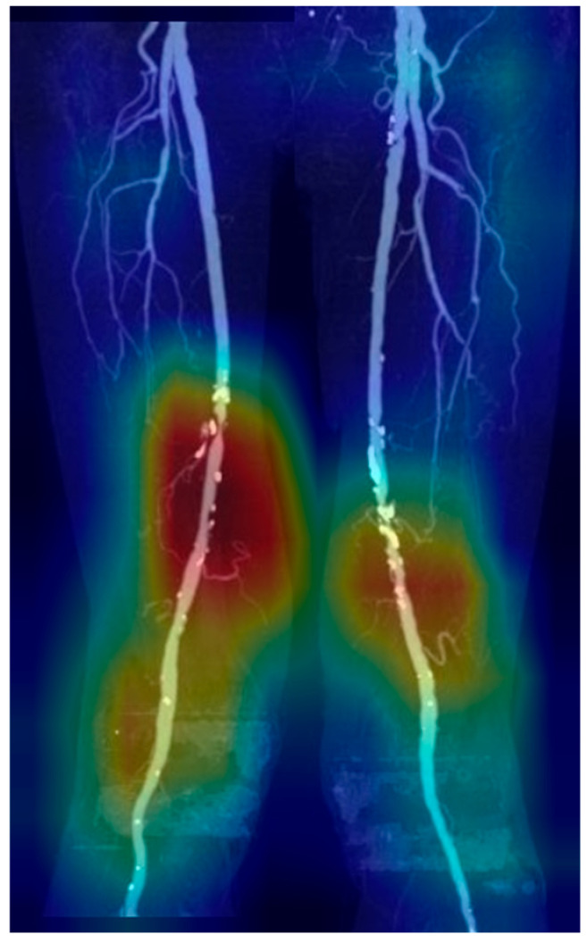
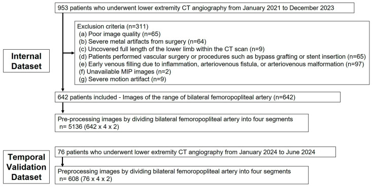
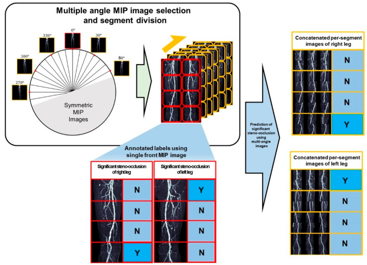
Background/Objectives: To develop and validate deep learning-based models for detecting significant steno-occlusion (SSO)-defined as luminal narrowing greater than 50%-of the femoropopliteal arteries using maximum intensity projection (MIP) images from lower extremity CT angiography (CTA). Methods: This retrospective study utilized MIP images of lower extremity CTA performed between January 2021 and December 2023 for internal model development. Deep learning-based models were developed sequentially to diagnose SSO: screening with single anteroposterior image, followed by four-segment rotational analysis that divided each femoropopliteal artery into four segments and incorporated multi-angle images. Given the cropped images and the shape of stenosis, models were trained to classify the presence of SSO. A temporal validation dataset comprised MIP images from lower extremity CTA performed between January and June 2024. Results: In total, 56,496 segment images from 642 patients (mean age: 68.2 ± 13.5 years; 472 men) were included in the internal dataset. In the single-image analysis, RDNet achieved the highest mean AUC of 0.886 for SSO detection. In the four-segment rotational analysis, RDNet also demonstrated the highest mean AUC, reaching 0.964 in both half-set and full-set approaches. While RDNet recorded the highest mean AUC, all other models showed improved AUCs as the number of input images increased (p < 0.05). In the temporal validation dataset, RDNet again achieved the highest mean AUC (0.959) in the half-set analysis. Conclusions: The deep learning-based model, particularly RDNet, demonstrated excellent performance in detecting SSO of peripheral arteries on MIP images from lower extremity CTA.
TomographyMedicine-Radiology, Nuclear Medicine and Imaging
CiteScore
2.70
自引率
10.50%
发文量
222
期刊介绍:
TomographyTM publishes basic (technical and pre-clinical) and clinical scientific articles which involve the advancement of imaging technologies. Tomography encompasses studies that use single or multiple imaging modalities including for example CT, US, PET, SPECT, MR and hyperpolarization technologies, as well as optical modalities (i.e. bioluminescence, photoacoustic, endomicroscopy, fiber optic imaging and optical computed tomography) in basic sciences, engineering, preclinical and clinical medicine.
Tomography also welcomes studies involving exploration and refinement of contrast mechanisms and image-derived metrics within and across modalities toward the development of novel imaging probes for image-based feedback and intervention. The use of imaging in biology and medicine provides unparalleled opportunities to noninvasively interrogate tissues to obtain real-time dynamic and quantitative information required for diagnosis and response to interventions and to follow evolving pathological conditions. As multi-modal studies and the complexities of imaging technologies themselves are ever increasing to provide advanced information to scientists and clinicians.
Tomography provides a unique publication venue allowing investigators the opportunity to more precisely communicate integrated findings related to the diverse and heterogeneous features associated with underlying anatomical, physiological, functional, metabolic and molecular genetic activities of normal and diseased tissue. Thus Tomography publishes peer-reviewed articles which involve the broad use of imaging of any tissue and disease type including both preclinical and clinical investigations. In addition, hardware/software along with chemical and molecular probe advances are welcome as they are deemed to significantly contribute towards the long-term goal of improving the overall impact of imaging on scientific and clinical discovery.

 求助内容:
求助内容: 应助结果提醒方式:


