D. О. Solovyevа, О. Y. Popadinets, I. S. Kolpashnikov, A. V. Altunina, V. A. Oleinikov
{"title":"Integration of Software for Nanotomography-Based 3D Tissue Reconstruction","authors":"D. О. Solovyevа, О. Y. Popadinets, I. S. Kolpashnikov, A. V. Altunina, V. A. Oleinikov","doi":"10.1134/S1068162024607146","DOIUrl":null,"url":null,"abstract":"<p><b>Objective:</b> To enhance three-dimensional (3D) reconstruction of cellular tissues and their components at the nano scale by integrating dedicated software tools into optical probe nanotomography (OPNT). <b>Methods:</b> We incorporated the 3D Slicer open source platform into the OPNT workflow to standardize, optimize, and automate volumetric reconstruction. Atomic force microscopy (AFM) and optical microscopy datasets were imported into 3D Slicer, aligned, segmented, and rendered to generate high-resolution 3D models. A fragment of an astrocyte was used as a test sample to validate reconstruction accuracy and workflow efficiency. <b>Results and Discussion:</b> The enhanced OPNT pipeline produced reliable and reproducible 3D reconstructions of complex biological structures. Integration of 3D Slicer enabled precise registration of multimodal images and flexible segmentation tools, resulting in detailed morphology of astrocytic processes. Quantitative evaluation demonstrated improved spatial resolution and reduced user bias compared to manual reconstruction methods. The platform’s modularity facilitates adaptation to varied microscopy datasets and reconstruction tasks. <b>Conclusions:</b> The proposed methodology provides a robust, high-precision platform for 3D reconstruction of cellular and subcellular structures. It holds significant potential for specialized applications such as synaptic environment analysis of individual neurons and broad use in biomedical research and materials science.</p>","PeriodicalId":758,"journal":{"name":"Russian Journal of Bioorganic Chemistry","volume":"51 4","pages":"1579 - 1585"},"PeriodicalIF":1.7000,"publicationDate":"2025-07-28","publicationTypes":"Journal Article","fieldsOfStudy":null,"isOpenAccess":false,"openAccessPdf":"","citationCount":"0","resultStr":null,"platform":"Semanticscholar","paperid":null,"PeriodicalName":"Russian Journal of Bioorganic Chemistry","FirstCategoryId":"92","ListUrlMain":"https://link.springer.com/article/10.1134/S1068162024607146","RegionNum":4,"RegionCategory":"化学","ArticlePicture":[],"TitleCN":null,"AbstractTextCN":null,"PMCID":null,"EPubDate":"","PubModel":"","JCR":"Q4","JCRName":"BIOCHEMISTRY & MOLECULAR BIOLOGY","Score":null,"Total":0}
引用次数: 0
Abstract
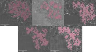
Objective: To enhance three-dimensional (3D) reconstruction of cellular tissues and their components at the nano scale by integrating dedicated software tools into optical probe nanotomography (OPNT). Methods: We incorporated the 3D Slicer open source platform into the OPNT workflow to standardize, optimize, and automate volumetric reconstruction. Atomic force microscopy (AFM) and optical microscopy datasets were imported into 3D Slicer, aligned, segmented, and rendered to generate high-resolution 3D models. A fragment of an astrocyte was used as a test sample to validate reconstruction accuracy and workflow efficiency. Results and Discussion: The enhanced OPNT pipeline produced reliable and reproducible 3D reconstructions of complex biological structures. Integration of 3D Slicer enabled precise registration of multimodal images and flexible segmentation tools, resulting in detailed morphology of astrocytic processes. Quantitative evaluation demonstrated improved spatial resolution and reduced user bias compared to manual reconstruction methods. The platform’s modularity facilitates adaptation to varied microscopy datasets and reconstruction tasks. Conclusions: The proposed methodology provides a robust, high-precision platform for 3D reconstruction of cellular and subcellular structures. It holds significant potential for specialized applications such as synaptic environment analysis of individual neurons and broad use in biomedical research and materials science.
期刊介绍:
Russian Journal of Bioorganic Chemistry publishes reviews and original experimental and theoretical studies on the structure, function, structure–activity relationships, and synthesis of biopolymers, such as proteins, nucleic acids, polysaccharides, mixed biopolymers, and their complexes, and low-molecular-weight biologically active compounds (peptides, sugars, lipids, antibiotics, etc.). The journal also covers selected aspects of neuro- and immunochemistry, biotechnology, and ecology.
 求助内容:
求助内容: 应助结果提醒方式:


