Edie Benedito Caetano, Cristina Schmitt H Cavalheiro, Núbia Dos Reis Sampaio, Pedro Mariano Coelho, Luiz Angelo Vieira, Julio Cesar Gali
{"title":"ANATOMOTOPOGRAPHIC STUDY OF THE MARTIN-GRUBER ANASTOMOSIS.","authors":"Edie Benedito Caetano, Cristina Schmitt H Cavalheiro, Núbia Dos Reis Sampaio, Pedro Mariano Coelho, Luiz Angelo Vieira, Julio Cesar Gali","doi":"10.1590/1413-785220253305e290615","DOIUrl":null,"url":null,"abstract":"<p><strong>Objective: </strong>To create, through anatomical dissections, a map of the location of the Martin-Gruber anastomosis (MGA) in the forearms of cadavers.</p><p><strong>Methods: </strong>One hundred forearms from 50 adult cadavers were used in this study. Dissection was performed through a median incision in the forearm and distal third of the arm. Lines between the humeral epicondyles (interepicondylar) and between the styloid processes of the radius and ulna (interstyloidea) were used as reference points for the topographic location of the anastomoses, and the forearms were divided into proximal, middle and distal thirds.</p><p><strong>Results: </strong>MGA was present in 27 forearms (27%). In four limbs (14.8%) the nerve fascicles originated from the median nerve proximal to the interepicondylar line. In two limbs (7.4%), at the level of the interepicondylar line and, in 21 of these (77.7%), they were found distal to this line. In 17 limbs (62.9%), the anastomosis occurred in the proximal third of the forearm, in eight limbs (29.6%), the anastomosis occurred in the middle third of the forearm and, in two limbs (7.4%), the anastomosis occurred with the ulnar nerve it occurred in the distal third of the forearm.</p><p><strong>Conclusion: </strong>Despite the great variation in their location, most anastomoses were found distal to the interepicondylar line, especially in the proximal third of the forearm. <b><i>Level of Evidence IV; Case Series.</i></b></p>","PeriodicalId":55563,"journal":{"name":"Acta Ortopedica Brasileira","volume":"33 5","pages":"e290615"},"PeriodicalIF":0.6000,"publicationDate":"2025-09-22","publicationTypes":"Journal Article","fieldsOfStudy":null,"isOpenAccess":false,"openAccessPdf":"https://www.ncbi.nlm.nih.gov/pmc/articles/PMC12456898/pdf/","citationCount":"0","resultStr":null,"platform":"Semanticscholar","paperid":null,"PeriodicalName":"Acta Ortopedica Brasileira","FirstCategoryId":"3","ListUrlMain":"https://doi.org/10.1590/1413-785220253305e290615","RegionNum":4,"RegionCategory":"医学","ArticlePicture":[],"TitleCN":null,"AbstractTextCN":null,"PMCID":null,"EPubDate":"2025/1/1 0:00:00","PubModel":"eCollection","JCR":"Q4","JCRName":"ORTHOPEDICS","Score":null,"Total":0}
引用次数: 0
Abstract
Objective: To create, through anatomical dissections, a map of the location of the Martin-Gruber anastomosis (MGA) in the forearms of cadavers.
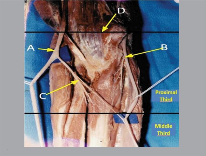
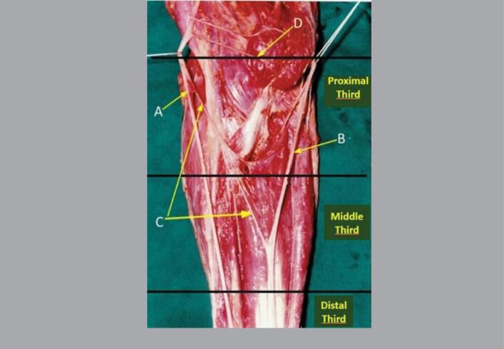
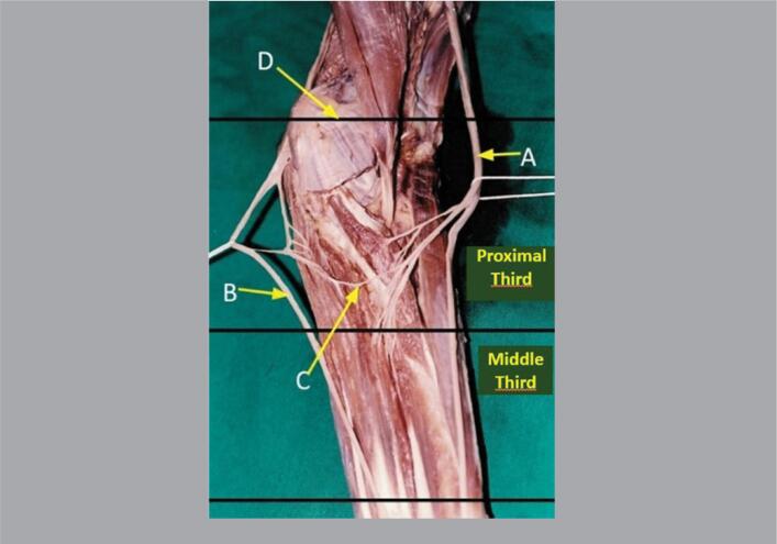
Methods: One hundred forearms from 50 adult cadavers were used in this study. Dissection was performed through a median incision in the forearm and distal third of the arm. Lines between the humeral epicondyles (interepicondylar) and between the styloid processes of the radius and ulna (interstyloidea) were used as reference points for the topographic location of the anastomoses, and the forearms were divided into proximal, middle and distal thirds.
Results: MGA was present in 27 forearms (27%). In four limbs (14.8%) the nerve fascicles originated from the median nerve proximal to the interepicondylar line. In two limbs (7.4%), at the level of the interepicondylar line and, in 21 of these (77.7%), they were found distal to this line. In 17 limbs (62.9%), the anastomosis occurred in the proximal third of the forearm, in eight limbs (29.6%), the anastomosis occurred in the middle third of the forearm and, in two limbs (7.4%), the anastomosis occurred with the ulnar nerve it occurred in the distal third of the forearm.
Conclusion: Despite the great variation in their location, most anastomoses were found distal to the interepicondylar line, especially in the proximal third of the forearm. Level of Evidence IV; Case Series.
期刊介绍:
A Revista Acta Ortopédica Brasileira, órgão oficial do Departamento de Ortopedia e Traumatologia da Faculdade de Medicina da Universidade de São Paulo (DOT/FMUSP), é publicada bimestralmente em seis edições ao ano (jan/fev, mar/abr, maio/jun, jul/ago, set/out e nov/dez) com versão em inglês disponível nos principais indexadores nacionais e internacionais e instituições de ensino do Brasil. Sendo hoje reconhecidamente uma importante contribuição para os especialistas da área com sua seriedade e árduo trabalho para as indexações já conquistadas.

 求助内容:
求助内容: 应助结果提醒方式:


