{"title":"Positive Relationships between Intraocular Pressure and Thyroid Function in Graves' Ophthalmopathy: A Three-Aspect Evidence Analysis.","authors":"Wenhua Zhang, Feng Zhang, Ziyi Zhu, Jiamin Cao, Wei Xiong","doi":"10.3349/ymj.2024.0468","DOIUrl":null,"url":null,"abstract":"<p><strong>Purpose: </strong>High intraocular pressure (IOP) is a frequent clinical manifestation of Graves' ophthalmopathy (GO), but little is known about the mechanism by which thyroid function alters IOP in GO.</p><p><strong>Materials and methods: </strong>The relationships between IOP and clinical manifestations of GO were determined by simple linear correlation analysis and the Kruskal-Wallis test. Factors significantly associated with changes in IOP were evaluated by multiple linear regression analysis. The relationships between IOP and thyroid function were verified using data from the National Health and Nutrition Examination Survey, and causative relationships between thyroid function and IOP were assessed using summary-data-based Mendelian randomization analysis. Gene expression was compared in patients with and without GO using analysis of a Gene Expression Omnibus dataset, and associated GO pathways were identified.</p><p><strong>Results: </strong>Clinical data were obtained from 392 hospitalizations of 270 patients, including 142 hospitalizations with increased IOP and 250 with normal IOP. IOP was linearly associated with GO duration, clinical activity score (CAS), and concentrations of free triiodothyronine (FT3), free thyroxine (FT4), and thyrotropin receptor antibodies. Multiple linear regression analysis yielded a model including CAS, FT3 concentration, and sex (r=0.46). Analysis of 3848 samples from the dataset showed that FT3 and FT4 levels differed between the increased and normal IOP groups. Increased expression of the <i>LOXL-AS1</i> gene was identified as a contributor to increased IOP, with <i>LOXL-AS1</i> expression increased in GO.</p><p><strong>Conclusion: </strong>Increased thyroid function is a risk factor for elevated IOP in patients with GO, with higher expression of <i>LOXL-AS1</i> in GO contributing to this effect.</p>","PeriodicalId":23765,"journal":{"name":"Yonsei Medical Journal","volume":"66 10","pages":"675-684"},"PeriodicalIF":2.8000,"publicationDate":"2025-10-01","publicationTypes":"Journal Article","fieldsOfStudy":null,"isOpenAccess":false,"openAccessPdf":"https://www.ncbi.nlm.nih.gov/pmc/articles/PMC12479196/pdf/","citationCount":"0","resultStr":null,"platform":"Semanticscholar","paperid":null,"PeriodicalName":"Yonsei Medical Journal","FirstCategoryId":"3","ListUrlMain":"https://doi.org/10.3349/ymj.2024.0468","RegionNum":4,"RegionCategory":"医学","ArticlePicture":[],"TitleCN":null,"AbstractTextCN":null,"PMCID":null,"EPubDate":"","PubModel":"","JCR":"Q1","JCRName":"MEDICINE, GENERAL & INTERNAL","Score":null,"Total":0}
引用次数: 0
Abstract
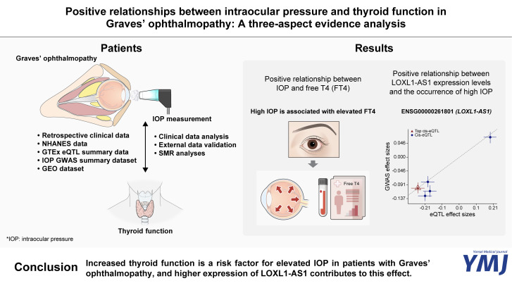
Purpose: High intraocular pressure (IOP) is a frequent clinical manifestation of Graves' ophthalmopathy (GO), but little is known about the mechanism by which thyroid function alters IOP in GO.
Materials and methods: The relationships between IOP and clinical manifestations of GO were determined by simple linear correlation analysis and the Kruskal-Wallis test. Factors significantly associated with changes in IOP were evaluated by multiple linear regression analysis. The relationships between IOP and thyroid function were verified using data from the National Health and Nutrition Examination Survey, and causative relationships between thyroid function and IOP were assessed using summary-data-based Mendelian randomization analysis. Gene expression was compared in patients with and without GO using analysis of a Gene Expression Omnibus dataset, and associated GO pathways were identified.
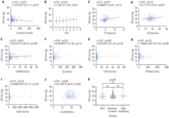
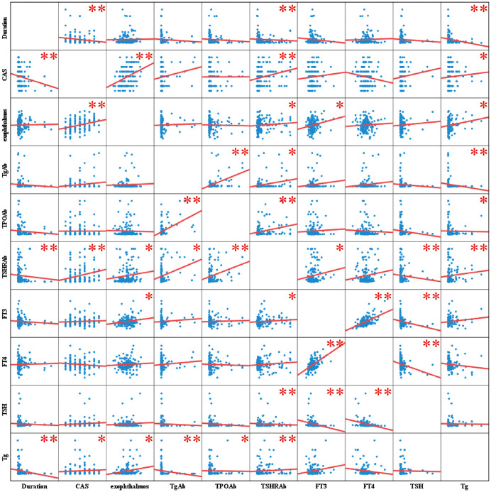
Results: Clinical data were obtained from 392 hospitalizations of 270 patients, including 142 hospitalizations with increased IOP and 250 with normal IOP. IOP was linearly associated with GO duration, clinical activity score (CAS), and concentrations of free triiodothyronine (FT3), free thyroxine (FT4), and thyrotropin receptor antibodies. Multiple linear regression analysis yielded a model including CAS, FT3 concentration, and sex (r=0.46). Analysis of 3848 samples from the dataset showed that FT3 and FT4 levels differed between the increased and normal IOP groups. Increased expression of the LOXL-AS1 gene was identified as a contributor to increased IOP, with LOXL-AS1 expression increased in GO.
Conclusion: Increased thyroid function is a risk factor for elevated IOP in patients with GO, with higher expression of LOXL-AS1 in GO contributing to this effect.
期刊介绍:
The goal of the Yonsei Medical Journal (YMJ) is to publish high quality manuscripts dedicated to clinical or basic research. Any authors affiliated with an accredited biomedical institution may submit manuscripts of original articles, review articles, case reports, brief communications, and letters to the Editor.
 求助内容:
求助内容: 应助结果提醒方式:


