{"title":"Conformational changes, excess area, and elasticity of the Piezo protein-membrane nanodome from coarse-grained and atomistic simulations.","authors":"Sneha Dixit, Frank Noé, Thomas R Weikl","doi":"10.7554/eLife.105138","DOIUrl":null,"url":null,"abstract":"<p><p>The mechanosensitive ion channels Piezo 1 and 2 induce a curved protein-membrane nanodome that flattens with increasing membrane tension γ. The tension-induced flattening of the nanodome is associated with Piezo activation and driven by the energy γΔA where ΔA is the excess area of the curved nanodome relative to its planar projected area. Based on extensive coarse-grained and atomistic simulations of membrane-embedded Piezo 1 and 2 proteins, we report here an excess area ΔA for the Piezo protein-membrane nanodome of about 40 nm<sup>2</sup> in tensionless membranes, and a half-maximal reduction of ΔA at tension values of about 3-4 mN/m, which is within the range of experimentally determined values for the half-maximal activation of Piezo 1. In line with recent experimental investigations of Piezo proteins in cell membranes and membrane vesicles, the membrane-embedded Piezo proteins adopt conformations in our simulations that are significantly less curved than the protein conformation in the detergent micelles of cryo-EM structures. An elasticity analysis of the nanodome shapes and protein conformations obtained from our simulations leads to an elastic model for Piezo activation that distinguishes the different energy components of the protein and the membrane in the tension-induced flattening of the nanodome. According to this model, the Piezo proteins resist flattening with a force constant of about 60 pN/nm.</p>","PeriodicalId":11640,"journal":{"name":"eLife","volume":"14 ","pages":""},"PeriodicalIF":6.4000,"publicationDate":"2025-09-24","publicationTypes":"Journal Article","fieldsOfStudy":null,"isOpenAccess":false,"openAccessPdf":"https://www.ncbi.nlm.nih.gov/pmc/articles/PMC12459952/pdf/","citationCount":"0","resultStr":null,"platform":"Semanticscholar","paperid":null,"PeriodicalName":"eLife","FirstCategoryId":"99","ListUrlMain":"https://doi.org/10.7554/eLife.105138","RegionNum":1,"RegionCategory":"生物学","ArticlePicture":[],"TitleCN":null,"AbstractTextCN":null,"PMCID":null,"EPubDate":"","PubModel":"","JCR":"Q1","JCRName":"BIOLOGY","Score":null,"Total":0}
引用次数: 0
Abstract
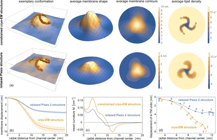
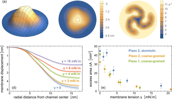
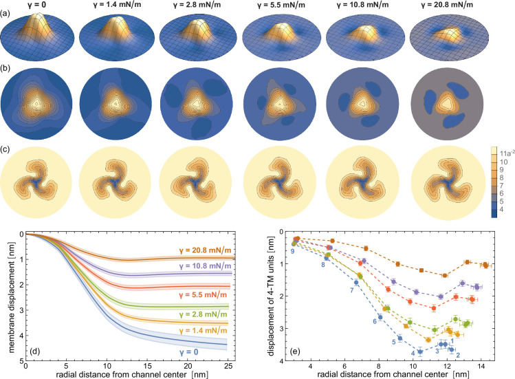
The mechanosensitive ion channels Piezo 1 and 2 induce a curved protein-membrane nanodome that flattens with increasing membrane tension γ. The tension-induced flattening of the nanodome is associated with Piezo activation and driven by the energy γΔA where ΔA is the excess area of the curved nanodome relative to its planar projected area. Based on extensive coarse-grained and atomistic simulations of membrane-embedded Piezo 1 and 2 proteins, we report here an excess area ΔA for the Piezo protein-membrane nanodome of about 40 nm2 in tensionless membranes, and a half-maximal reduction of ΔA at tension values of about 3-4 mN/m, which is within the range of experimentally determined values for the half-maximal activation of Piezo 1. In line with recent experimental investigations of Piezo proteins in cell membranes and membrane vesicles, the membrane-embedded Piezo proteins adopt conformations in our simulations that are significantly less curved than the protein conformation in the detergent micelles of cryo-EM structures. An elasticity analysis of the nanodome shapes and protein conformations obtained from our simulations leads to an elastic model for Piezo activation that distinguishes the different energy components of the protein and the membrane in the tension-induced flattening of the nanodome. According to this model, the Piezo proteins resist flattening with a force constant of about 60 pN/nm.
期刊介绍:
eLife is a distinguished, not-for-profit, peer-reviewed open access scientific journal that specializes in the fields of biomedical and life sciences. eLife is known for its selective publication process, which includes a variety of article types such as:
Research Articles: Detailed reports of original research findings.
Short Reports: Concise presentations of significant findings that do not warrant a full-length research article.
Tools and Resources: Descriptions of new tools, technologies, or resources that facilitate scientific research.
Research Advances: Brief reports on significant scientific advancements that have immediate implications for the field.
Scientific Correspondence: Short communications that comment on or provide additional information related to published articles.
Review Articles: Comprehensive overviews of a specific topic or field within the life sciences.
 求助内容:
求助内容: 应助结果提醒方式:


