Frederik Fuchs, Sebastian N Marschner, Jan Hofmaier, Maya Rottler, Indra Hadi, Sebastian H Maier, Tobias Greve, Adrien Holzgreve, Nathalie L Albert, Raphael Bodensohn, Claus Belka, Maximilian Niyazi, Franziska Walter
{"title":"SSTR PET/CT for skull base low-grade meningioma: a critical tool for accurate gross tumor volume delineation in radiotherapy?","authors":"Frederik Fuchs, Sebastian N Marschner, Jan Hofmaier, Maya Rottler, Indra Hadi, Sebastian H Maier, Tobias Greve, Adrien Holzgreve, Nathalie L Albert, Raphael Bodensohn, Claus Belka, Maximilian Niyazi, Franziska Walter","doi":"10.1186/s13014-025-02718-4","DOIUrl":null,"url":null,"abstract":"<p><strong>Background: </strong>Precise delineation of gross tumor volume (GTV) is fundamental for effective radiation therapy in low-grade skull base meningiomas. Magnetic resonance imaging (MRI) serves as the primary imaging tool but may not fully represent tumor extent. This study investigates the additional value of incorporating Somatostatin receptor (SSTR)-directed PET/CT in radiation therapy planning.</p><p><strong>Methods: </strong>A retrospective analysis was conducted with four experienced radiation oncologists contouring GTVs for skull base meningiomas using MRI alone (GTV_MRI), PET/CT alone (GTV_PET/CT), and both modalities combined (GTV_ALL). Consensus ground truth volumes were generated for each modality through a STAPLE algorithm. Agreement between modalities, excluding observer variability, was assessed using statistical metrics including Dice Similarity Coefficient (DSC), Jaccard Index (JCI), Hausdorff distance (HD95), Geographical Miss Index (GMI), sensitivity, and kappa statistics.</p><p><strong>Results: </strong>The study included 25 patients (15 females, 10 males; median age 56 years (range: 23-74 years), with 96% achieving local control post-radiotherapy over a median follow-up of 64 months (range: 28-135 months). Substantial interobserver agreement was observed, with median kappa values of 0.74 for GTV_MRI, 0.68 for GTV_PET/CT, and 0.77 for GTV_ALL. Median consensus volumes were 6.65 cc (MRI<sub>STAPLE</sub>), 7.21 cc (PET<sub>STAPLE</sub>), and 6.73 cc (ALL<sub>STAPLE</sub>). The median GMI for MRI<sub>STAPLE</sub> compared to ALL<sub>STAPLE</sub> was 0.18 (IQR: 0.11-0.39), and 0.21 (IQR: 0.15-0.28) for PET<sub>STAPLE</sub> compared to ALL<sub>STAPLE</sub>. The DSC indicated the lowest concordance between MRI<sub>STAPLE</sub> and PET<sub>STAPLE</sub> with a median of 0.75 (IQR: 0.59-0.82), followed by PET<sub>STAPLE</sub> versus ALL<sub>STAPLE</sub> with a median DSC of 0.84 (IQR: 0.79-0.89), and MRI<sub>STAPLE</sub> versus ALL<sub>STAPLE</sub> with a median DSC of 0.89 (IQR: 0.76-0.92). The integration of PET/CT with MRI significantly enhanced concordance metrics.</p><p><strong>Conclusion: </strong>Combining MRI and PET/CT improves GTV delineation in low-grade skull base meningiomas, as PET/CT can reveal regions missed by MRI, which may slightly underestimate tumor size. This multimodal imaging approach enhances consensus and supports its role in radiotherapy planning. Standardized protocols and technical integration remain key future goals.</p>","PeriodicalId":49639,"journal":{"name":"Radiation Oncology","volume":"20 1","pages":"142"},"PeriodicalIF":3.3000,"publicationDate":"2025-09-22","publicationTypes":"Journal Article","fieldsOfStudy":null,"isOpenAccess":false,"openAccessPdf":"https://www.ncbi.nlm.nih.gov/pmc/articles/PMC12455788/pdf/","citationCount":"0","resultStr":null,"platform":"Semanticscholar","paperid":null,"PeriodicalName":"Radiation Oncology","FirstCategoryId":"3","ListUrlMain":"https://doi.org/10.1186/s13014-025-02718-4","RegionNum":2,"RegionCategory":"医学","ArticlePicture":[],"TitleCN":null,"AbstractTextCN":null,"PMCID":null,"EPubDate":"","PubModel":"","JCR":"Q2","JCRName":"ONCOLOGY","Score":null,"Total":0}
引用次数: 0
Abstract
Background: Precise delineation of gross tumor volume (GTV) is fundamental for effective radiation therapy in low-grade skull base meningiomas. Magnetic resonance imaging (MRI) serves as the primary imaging tool but may not fully represent tumor extent. This study investigates the additional value of incorporating Somatostatin receptor (SSTR)-directed PET/CT in radiation therapy planning.
Methods: A retrospective analysis was conducted with four experienced radiation oncologists contouring GTVs for skull base meningiomas using MRI alone (GTV_MRI), PET/CT alone (GTV_PET/CT), and both modalities combined (GTV_ALL). Consensus ground truth volumes were generated for each modality through a STAPLE algorithm. Agreement between modalities, excluding observer variability, was assessed using statistical metrics including Dice Similarity Coefficient (DSC), Jaccard Index (JCI), Hausdorff distance (HD95), Geographical Miss Index (GMI), sensitivity, and kappa statistics.
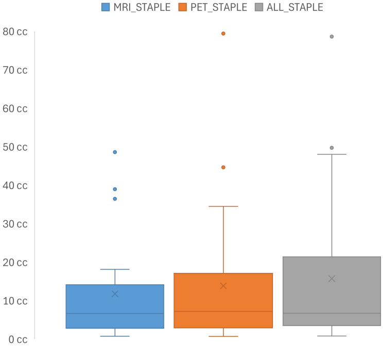
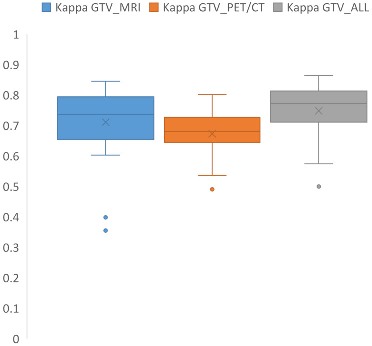
Results: The study included 25 patients (15 females, 10 males; median age 56 years (range: 23-74 years), with 96% achieving local control post-radiotherapy over a median follow-up of 64 months (range: 28-135 months). Substantial interobserver agreement was observed, with median kappa values of 0.74 for GTV_MRI, 0.68 for GTV_PET/CT, and 0.77 for GTV_ALL. Median consensus volumes were 6.65 cc (MRISTAPLE), 7.21 cc (PETSTAPLE), and 6.73 cc (ALLSTAPLE). The median GMI for MRISTAPLE compared to ALLSTAPLE was 0.18 (IQR: 0.11-0.39), and 0.21 (IQR: 0.15-0.28) for PETSTAPLE compared to ALLSTAPLE. The DSC indicated the lowest concordance between MRISTAPLE and PETSTAPLE with a median of 0.75 (IQR: 0.59-0.82), followed by PETSTAPLE versus ALLSTAPLE with a median DSC of 0.84 (IQR: 0.79-0.89), and MRISTAPLE versus ALLSTAPLE with a median DSC of 0.89 (IQR: 0.76-0.92). The integration of PET/CT with MRI significantly enhanced concordance metrics.
Conclusion: Combining MRI and PET/CT improves GTV delineation in low-grade skull base meningiomas, as PET/CT can reveal regions missed by MRI, which may slightly underestimate tumor size. This multimodal imaging approach enhances consensus and supports its role in radiotherapy planning. Standardized protocols and technical integration remain key future goals.
Radiation OncologyONCOLOGY-RADIOLOGY, NUCLEAR MEDICINE & MEDICAL IMAGING
CiteScore
6.50
自引率
2.80%
发文量
181
审稿时长
3-6 weeks
期刊介绍:
Radiation Oncology encompasses all aspects of research that impacts on the treatment of cancer using radiation. It publishes findings in molecular and cellular radiation biology, radiation physics, radiation technology, and clinical oncology.
 求助内容:
求助内容: 应助结果提醒方式:


