CT-Based 2.5D Deep Learning-Multi-Instance Learning for Predicting Early Recurrence of Hepatocellular Carcinoma and Correlating with Recurrence-Related Pathological Indicators.
Yongyi Cen, Haiyang Nong, Dehui Du, Yingning Wu, Jianpeng Chen, Zhaolin Pan, Yin Huang, Ke Ding, Deyou Huang
{"title":"CT-Based 2.5D Deep Learning-Multi-Instance Learning for Predicting Early Recurrence of Hepatocellular Carcinoma and Correlating with Recurrence-Related Pathological Indicators.","authors":"Yongyi Cen, Haiyang Nong, Dehui Du, Yingning Wu, Jianpeng Chen, Zhaolin Pan, Yin Huang, Ke Ding, Deyou Huang","doi":"10.2147/JHC.S541402","DOIUrl":null,"url":null,"abstract":"<p><strong>Purpose: </strong>This study aims to evaluate the advantages of the 2.5D deep learning-multi-instance learning (2.5D DL-MIL) model, based on CT arterial phase images, in predicting early recurrence (ER) of hepatocellular carcinoma (HCC) and examining the biological significance of MIL features.</p><p><strong>Patients and methods: </strong>A total of 191 HCC patients were retrospectively included and categorized into ER (n=79) and non-early recurrence (NER, n=112) groups based on postoperative follow-up results. The patients were randomly divided to the training set (n=133) and validation set (n=58) in a 7:3 ratio. The predictive capabilities of the 2.5D DL-MIL model, Radiomics model, and Clinical model for ER of HCC were constructed and compared using CT arterial phase and clinical data. SHAP analysis was used to evaluate the contribution of MIL features in the model, and further analysis was conducted on the correlation between MIL features and microvascular invasion (MVI), Ki-67 expression, and pathological grading.</p><p><strong>Results: </strong>The area under the curve (AUC) for the 2.5D DL-MIL model in the validation set was 0.840, surpassing that of the Radiomics model (AUC = 0.678, P = 0.047) and the Clinical model (AUC = 0.598, P = 0.009). Decision curve analyses indicated superior clinical utility for the 2.5D DL-MIL model. SHAP analysis revealed that bag-of-words features (eg, BoW_02 and BoW_09) were key contributors to the 2.5D DL-MIL model. Correlation analysis demonstrated that BoW_01, BoW_02, BoW_09, and BoW_1 were significantly correlated with MVI grade and Ki-67 expression (P < 0.05).</p><p><strong>Conclusion: </strong>The 2.5D DL-MIL model demonstrates significant value in predicting ER of HCC, with its MIL features exhibiting strong associations with tumor invasiveness and proliferative activity.</p>","PeriodicalId":15906,"journal":{"name":"Journal of Hepatocellular Carcinoma","volume":"12 ","pages":"2095-2108"},"PeriodicalIF":3.4000,"publicationDate":"2025-09-17","publicationTypes":"Journal Article","fieldsOfStudy":null,"isOpenAccess":false,"openAccessPdf":"https://www.ncbi.nlm.nih.gov/pmc/articles/PMC12450382/pdf/","citationCount":"0","resultStr":null,"platform":"Semanticscholar","paperid":null,"PeriodicalName":"Journal of Hepatocellular Carcinoma","FirstCategoryId":"3","ListUrlMain":"https://doi.org/10.2147/JHC.S541402","RegionNum":3,"RegionCategory":"医学","ArticlePicture":[],"TitleCN":null,"AbstractTextCN":null,"PMCID":null,"EPubDate":"2025/1/1 0:00:00","PubModel":"eCollection","JCR":"Q2","JCRName":"ONCOLOGY","Score":null,"Total":0}
引用次数: 0
Abstract
Purpose: This study aims to evaluate the advantages of the 2.5D deep learning-multi-instance learning (2.5D DL-MIL) model, based on CT arterial phase images, in predicting early recurrence (ER) of hepatocellular carcinoma (HCC) and examining the biological significance of MIL features.
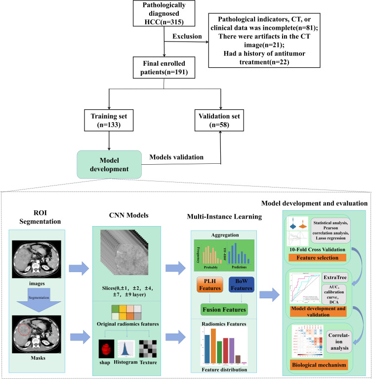
Patients and methods: A total of 191 HCC patients were retrospectively included and categorized into ER (n=79) and non-early recurrence (NER, n=112) groups based on postoperative follow-up results. The patients were randomly divided to the training set (n=133) and validation set (n=58) in a 7:3 ratio. The predictive capabilities of the 2.5D DL-MIL model, Radiomics model, and Clinical model for ER of HCC were constructed and compared using CT arterial phase and clinical data. SHAP analysis was used to evaluate the contribution of MIL features in the model, and further analysis was conducted on the correlation between MIL features and microvascular invasion (MVI), Ki-67 expression, and pathological grading.
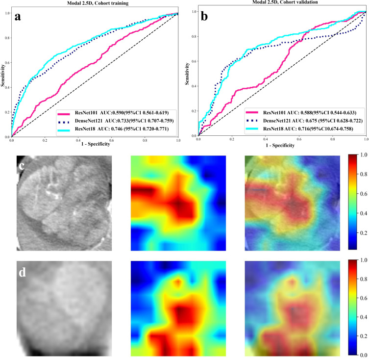
Results: The area under the curve (AUC) for the 2.5D DL-MIL model in the validation set was 0.840, surpassing that of the Radiomics model (AUC = 0.678, P = 0.047) and the Clinical model (AUC = 0.598, P = 0.009). Decision curve analyses indicated superior clinical utility for the 2.5D DL-MIL model. SHAP analysis revealed that bag-of-words features (eg, BoW_02 and BoW_09) were key contributors to the 2.5D DL-MIL model. Correlation analysis demonstrated that BoW_01, BoW_02, BoW_09, and BoW_1 were significantly correlated with MVI grade and Ki-67 expression (P < 0.05).
Conclusion: The 2.5D DL-MIL model demonstrates significant value in predicting ER of HCC, with its MIL features exhibiting strong associations with tumor invasiveness and proliferative activity.

 求助内容:
求助内容: 应助结果提醒方式:


