{"title":"Secondary aneurysmal bone cyst arising from polyostotic craniofacial fibrous dysplasia.","authors":"Niket Patel, Kiran Kumar Sailagundla, Essaki Rajulu, Shanmuga Jayanthan, Ganesh Rajagopal, Harpreet Singh Grewal","doi":"10.1093/bjrcr/uaaf047","DOIUrl":null,"url":null,"abstract":"<p><p>Fibrous dysplasia (FD) is a benign condition affecting osteoblasts, which fail to undergo proper differentiation and maturation, resulting in the replacement of normal osteoid matrix with ground glass fibrous tissue. Aneurysmal bone cyst (ABC) is a benign, expansile, lytic lesion characterized by multiple blood-filled cystic cavities containing haemorrhagic products at varying stages. Secondary ABC arising from craniofacial FD is extremely rare. To date, only 10 cases have been reported in the literature. This report highlights the clinical presentation, imaging findings, and histopathological confirmation of a secondary ABC in a patient with polyostotic craniofacial FD.</p>","PeriodicalId":45216,"journal":{"name":"BJR Case Reports","volume":"11 5","pages":"uaaf047"},"PeriodicalIF":0.5000,"publicationDate":"2025-09-09","publicationTypes":"Journal Article","fieldsOfStudy":null,"isOpenAccess":false,"openAccessPdf":"https://www.ncbi.nlm.nih.gov/pmc/articles/PMC12449245/pdf/","citationCount":"0","resultStr":null,"platform":"Semanticscholar","paperid":null,"PeriodicalName":"BJR Case Reports","FirstCategoryId":"1085","ListUrlMain":"https://doi.org/10.1093/bjrcr/uaaf047","RegionNum":0,"RegionCategory":null,"ArticlePicture":[],"TitleCN":null,"AbstractTextCN":null,"PMCID":null,"EPubDate":"2025/9/1 0:00:00","PubModel":"eCollection","JCR":"Q4","JCRName":"RADIOLOGY, NUCLEAR MEDICINE & MEDICAL IMAGING","Score":null,"Total":0}
引用次数: 0
Abstract
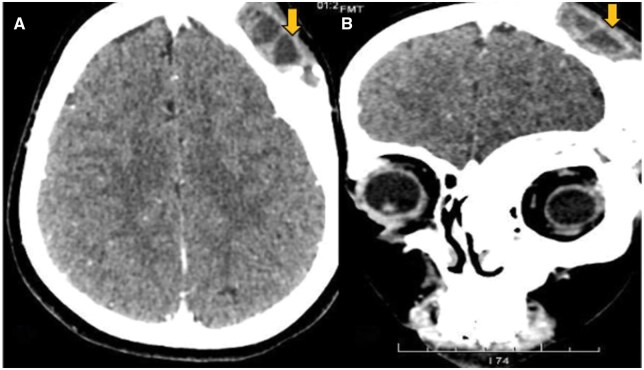
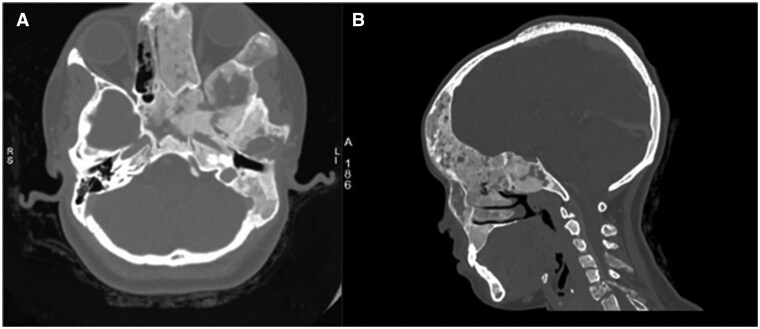
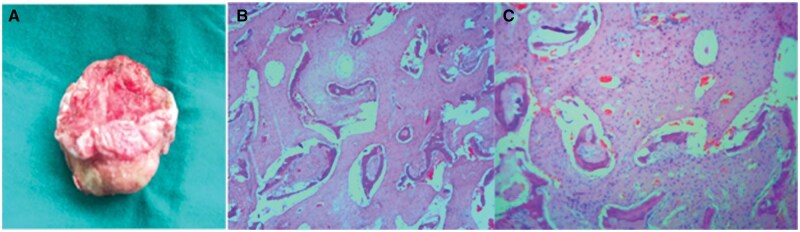
Fibrous dysplasia (FD) is a benign condition affecting osteoblasts, which fail to undergo proper differentiation and maturation, resulting in the replacement of normal osteoid matrix with ground glass fibrous tissue. Aneurysmal bone cyst (ABC) is a benign, expansile, lytic lesion characterized by multiple blood-filled cystic cavities containing haemorrhagic products at varying stages. Secondary ABC arising from craniofacial FD is extremely rare. To date, only 10 cases have been reported in the literature. This report highlights the clinical presentation, imaging findings, and histopathological confirmation of a secondary ABC in a patient with polyostotic craniofacial FD.

 求助内容:
求助内容: 应助结果提醒方式:


