Ayentika Sen, Aruna K. Mora, Soumitra Kundu, Sukhendu Nath
{"title":"Role of Ion-Pair and H-Bonding in Colorimetric Detection of Insulin Fibrils with Picomolar Sensitivity","authors":"Ayentika Sen, Aruna K. Mora, Soumitra Kundu, Sukhendu Nath","doi":"10.1002/cptc.202400329","DOIUrl":null,"url":null,"abstract":"<p>Ultrasensitive detection of amyloid fibril, a biomarker of several neurological diseases, has been achieved using a hemicyanine dye. Amyloid fibril induces a remarkable change in the absorption spectral position (~115 nm) of the perchlorate salt of the probe leading to a naked-eye detection of these neurotoxic protein aggregates. Such a large shift in the absorption spectra has been utilised to develop a simple and cost effective paper based detection of insulin aggregates with a limit of detection (LOD) of less than 2 pM. Besides the changes in the absorption spectra, the emission intensity of the probe shows almost two orders of magnitude increase in the presence of insulin amyloid fibrils. Such dual sensing properties of the present hemicyanine molecule make it a noble probe for amyloid fibrils. Our detailed investigation on the fundamental mechanism responsible for the fibril-induced spectral changes in the dye has established the role of amyloid induced dissociation of ion-pair and solute-solvent hydrogen bonding in the colorimetric detection of amyloid fibrils. Quantum chemical calculations and blind molecular docking studies have also been performed to strengthen our experimental observations. This result will open up a new frontier in the field of amyloid probes based on the unique mechanism proposed in this work.</p>","PeriodicalId":10108,"journal":{"name":"ChemPhotoChem","volume":"9 9","pages":""},"PeriodicalIF":3.0000,"publicationDate":"2025-02-18","publicationTypes":"Journal Article","fieldsOfStudy":null,"isOpenAccess":false,"openAccessPdf":"https://chemistry-europe.onlinelibrary.wiley.com/doi/epdf/10.1002/cptc.202400329","citationCount":"0","resultStr":null,"platform":"Semanticscholar","paperid":null,"PeriodicalName":"ChemPhotoChem","FirstCategoryId":"92","ListUrlMain":"https://chemistry-europe.onlinelibrary.wiley.com/doi/10.1002/cptc.202400329","RegionNum":4,"RegionCategory":"化学","ArticlePicture":[],"TitleCN":null,"AbstractTextCN":null,"PMCID":null,"EPubDate":"","PubModel":"","JCR":"Q3","JCRName":"CHEMISTRY, PHYSICAL","Score":null,"Total":0}
引用次数: 0
Abstract
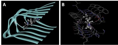
Ultrasensitive detection of amyloid fibril, a biomarker of several neurological diseases, has been achieved using a hemicyanine dye. Amyloid fibril induces a remarkable change in the absorption spectral position (~115 nm) of the perchlorate salt of the probe leading to a naked-eye detection of these neurotoxic protein aggregates. Such a large shift in the absorption spectra has been utilised to develop a simple and cost effective paper based detection of insulin aggregates with a limit of detection (LOD) of less than 2 pM. Besides the changes in the absorption spectra, the emission intensity of the probe shows almost two orders of magnitude increase in the presence of insulin amyloid fibrils. Such dual sensing properties of the present hemicyanine molecule make it a noble probe for amyloid fibrils. Our detailed investigation on the fundamental mechanism responsible for the fibril-induced spectral changes in the dye has established the role of amyloid induced dissociation of ion-pair and solute-solvent hydrogen bonding in the colorimetric detection of amyloid fibrils. Quantum chemical calculations and blind molecular docking studies have also been performed to strengthen our experimental observations. This result will open up a new frontier in the field of amyloid probes based on the unique mechanism proposed in this work.
ChemPhotoChemChemistry-Physical and Theoretical Chemistry
CiteScore
5.80
自引率
5.40%
发文量
165
期刊介绍:
Light plays a crucial role in natural processes and leads to exciting phenomena in molecules and materials. ChemPhotoChem welcomes exceptional international research in the entire scope of pure and applied photochemistry, photobiology, and photophysics. Our thorough editorial practices aid us in publishing authoritative research fast. We support the photochemistry community to be a leading light in science.
We understand the huge pressures the scientific community is facing every day and we want to support you. Chemistry Europe is an association of 16 chemical societies from 15 European countries. Run by chemists, for chemists—we evaluate, publish, disseminate, and amplify the scientific excellence of chemistry researchers from around the globe.


 求助内容:
求助内容: 应助结果提醒方式:


