Alexander Weiss, Alex Florin Meissner, Fanni Annamária Boros, Martin Regensburger, Regina Verena Taudte, Andreu Matamoros-Angles, Philipp Arnold, Friederike Zunke
{"title":"Transmission Electron Microscopy-based characterization of Extracellular Vesicles from plasma and serum from Parkinson´s Disease patients.","authors":"Alexander Weiss, Alex Florin Meissner, Fanni Annamária Boros, Martin Regensburger, Regina Verena Taudte, Andreu Matamoros-Angles, Philipp Arnold, Friederike Zunke","doi":"10.1186/s12964-025-02383-w","DOIUrl":null,"url":null,"abstract":"<p><p>Parkinson's disease is a neurodegenerative disorder with no curative treatment option and objective biomarker profile. Extracellular Vesicles (EVs) are membrane-enclosed biological nanoparticles released from all cells of the human body. In this pilot study we compare plasma- and serum-derived EVs from Parkinson's disease (PD) patients and healthy controls (HC) utilizing a precipitation-based method. Additionally, we employ an L1CAM antibody to selectively enrich for L1CAM-positive EVs from the total plasma-/serum-derived EV fractions. Successful EV enrichment was shown in western blot experiments for CD63 and for L1CAM as well as in metabolomic analysis for a HC sample. In a side-by-side quantification,. which we based on transmission electron microscopic images from negative stain samples, we identify small but significant differences between EV diameter from PD patients and HC. To streamline the quantification process, we introduce an ImageJ-based computer algorithm for (semi-)automated quantification of EVs from negative stain electron micrographs. We observe that this (semi-)automated quantification determines a smaller diameter than manual quantification. However, the difference between PD and HC group is systematic and reveals the same relative differences calculated from manually measured total plasma-derived EV particles. In this pilot study, we introduce a new workflow implemented into an ImageJ plugin enabling to determine differences in EV size within TEM images. For our data set of plasma-derived EVs from PD patients and HC, we find small, yet consistent differences. We feel that this study contributes to the search of a clinical biomarker for PD.</p>","PeriodicalId":55268,"journal":{"name":"Cell Communication and Signaling","volume":"23 1","pages":"395"},"PeriodicalIF":8.2000,"publicationDate":"2025-09-18","publicationTypes":"Journal Article","fieldsOfStudy":null,"isOpenAccess":false,"openAccessPdf":"https://www.ncbi.nlm.nih.gov/pmc/articles/PMC12445024/pdf/","citationCount":"0","resultStr":null,"platform":"Semanticscholar","paperid":null,"PeriodicalName":"Cell Communication and Signaling","FirstCategoryId":"99","ListUrlMain":"https://doi.org/10.1186/s12964-025-02383-w","RegionNum":2,"RegionCategory":"生物学","ArticlePicture":[],"TitleCN":null,"AbstractTextCN":null,"PMCID":null,"EPubDate":"","PubModel":"","JCR":"Q1","JCRName":"CELL BIOLOGY","Score":null,"Total":0}
引用次数: 0
Abstract
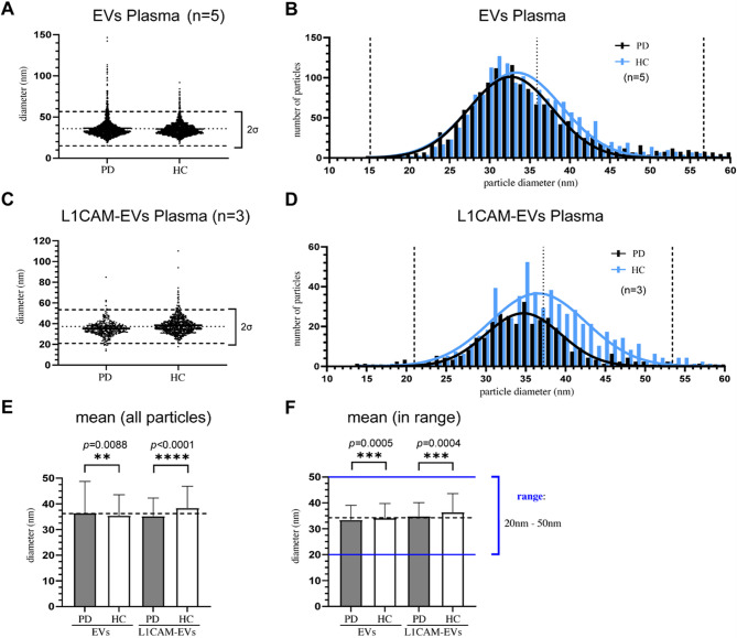
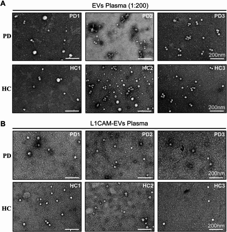
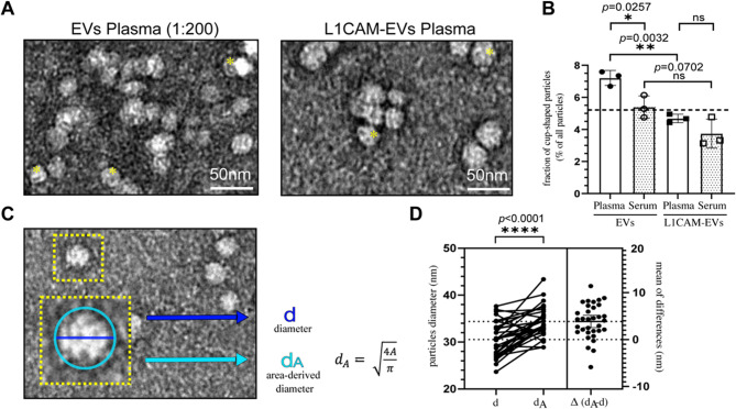
Parkinson's disease is a neurodegenerative disorder with no curative treatment option and objective biomarker profile. Extracellular Vesicles (EVs) are membrane-enclosed biological nanoparticles released from all cells of the human body. In this pilot study we compare plasma- and serum-derived EVs from Parkinson's disease (PD) patients and healthy controls (HC) utilizing a precipitation-based method. Additionally, we employ an L1CAM antibody to selectively enrich for L1CAM-positive EVs from the total plasma-/serum-derived EV fractions. Successful EV enrichment was shown in western blot experiments for CD63 and for L1CAM as well as in metabolomic analysis for a HC sample. In a side-by-side quantification,. which we based on transmission electron microscopic images from negative stain samples, we identify small but significant differences between EV diameter from PD patients and HC. To streamline the quantification process, we introduce an ImageJ-based computer algorithm for (semi-)automated quantification of EVs from negative stain electron micrographs. We observe that this (semi-)automated quantification determines a smaller diameter than manual quantification. However, the difference between PD and HC group is systematic and reveals the same relative differences calculated from manually measured total plasma-derived EV particles. In this pilot study, we introduce a new workflow implemented into an ImageJ plugin enabling to determine differences in EV size within TEM images. For our data set of plasma-derived EVs from PD patients and HC, we find small, yet consistent differences. We feel that this study contributes to the search of a clinical biomarker for PD.
期刊介绍:
Cell Communication and Signaling (CCS) is a peer-reviewed, open-access scientific journal that focuses on cellular signaling pathways in both normal and pathological conditions. It publishes original research, reviews, and commentaries, welcoming studies that utilize molecular, morphological, biochemical, structural, and cell biology approaches. CCS also encourages interdisciplinary work and innovative models, including in silico, in vitro, and in vivo approaches, to facilitate investigations of cell signaling pathways, networks, and behavior.
Starting from January 2019, CCS is proud to announce its affiliation with the International Cell Death Society. The journal now encourages submissions covering all aspects of cell death, including apoptotic and non-apoptotic mechanisms, cell death in model systems, autophagy, clearance of dying cells, and the immunological and pathological consequences of dying cells in the tissue microenvironment.

 求助内容:
求助内容: 应助结果提醒方式:


