Luis Javier Cajas Santana, Satiago Cuero, Gabriela Guerrero, Mayelin Ceballos, María Carolina Torres Villarreal
{"title":"Correlation of Pleural and Pulmonary Ultrasound with the Severity of Autoimmune Interstitial Lung Disease.","authors":"Luis Javier Cajas Santana, Satiago Cuero, Gabriela Guerrero, Mayelin Ceballos, María Carolina Torres Villarreal","doi":"10.5152/ArchRheumatol.2025.10999","DOIUrl":null,"url":null,"abstract":"<p><p>Background/Aims: Interstitial lung disease is one of the most important complications in autoimmune diseases. The extent of involvement in tomography is crucial for therapeutic decision-making. Pleuropulmonary ultrasound is helpful in screening and correlates with severity. Using the Goh method, the objective is to analyze the correlation between ultrasound findings and the quantified extent on high-resolution computed tomography (HRCT). Materials and Methods: The HRCT images of patients with rheumatoid arthritis or systemic sclerosis were analyzed using the Goh method. The data were compared with the number of B-lines and ultrasound pleural abnormalities. The correlation was determined using Spearman's Rho statistic, and receiver operating characteristic curve analysis was performed. The sensitivity, specificity, and cutoff points were calculated for each ultrasound finding to detect severe disease. Results: A total of 71 patients were included. Almost half of the patients (56%) were involved in less than 5% of extent in HRCT; an average disease extent was 11% for the whole population. The correlation (Rho) between the extent and the total B-line count was 0.58 and 0.61 (P < .001), and for pleural abnormalities, 0.60 and 0.59 (P < .001) in linear and convex images, correspondingly. The areas under the curve were high for both ultrasound abnormalities and in both forms of images, consistently exceeding 0.7. Regarding the cutoff values, a number greater than 20 B-lines has a specificity close to 90% for detecting extensive disease, as well as 7 or more pleural abnormalities. Conclusion: The count of B-lines and the number of pleural abnormalities on lung ultrasound correlate well with the extent of the disease and can help determine its severity.</p>","PeriodicalId":93884,"journal":{"name":"Archives of rheumatology","volume":"40 3","pages":"308-314"},"PeriodicalIF":1.1000,"publicationDate":"2025-09-01","publicationTypes":"Journal Article","fieldsOfStudy":null,"isOpenAccess":false,"openAccessPdf":"https://www.ncbi.nlm.nih.gov/pmc/articles/PMC12502842/pdf/","citationCount":"0","resultStr":null,"platform":"Semanticscholar","paperid":null,"PeriodicalName":"Archives of rheumatology","FirstCategoryId":"1085","ListUrlMain":"https://doi.org/10.5152/ArchRheumatol.2025.10999","RegionNum":0,"RegionCategory":null,"ArticlePicture":[],"TitleCN":null,"AbstractTextCN":null,"PMCID":null,"EPubDate":"","PubModel":"","JCR":"Q4","JCRName":"RHEUMATOLOGY","Score":null,"Total":0}
引用次数: 0
Abstract
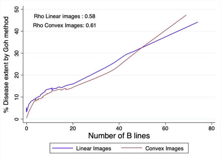
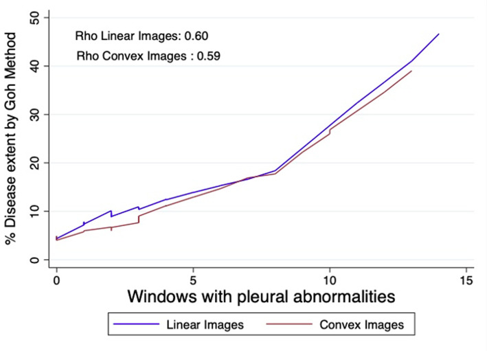
Background/Aims: Interstitial lung disease is one of the most important complications in autoimmune diseases. The extent of involvement in tomography is crucial for therapeutic decision-making. Pleuropulmonary ultrasound is helpful in screening and correlates with severity. Using the Goh method, the objective is to analyze the correlation between ultrasound findings and the quantified extent on high-resolution computed tomography (HRCT). Materials and Methods: The HRCT images of patients with rheumatoid arthritis or systemic sclerosis were analyzed using the Goh method. The data were compared with the number of B-lines and ultrasound pleural abnormalities. The correlation was determined using Spearman's Rho statistic, and receiver operating characteristic curve analysis was performed. The sensitivity, specificity, and cutoff points were calculated for each ultrasound finding to detect severe disease. Results: A total of 71 patients were included. Almost half of the patients (56%) were involved in less than 5% of extent in HRCT; an average disease extent was 11% for the whole population. The correlation (Rho) between the extent and the total B-line count was 0.58 and 0.61 (P < .001), and for pleural abnormalities, 0.60 and 0.59 (P < .001) in linear and convex images, correspondingly. The areas under the curve were high for both ultrasound abnormalities and in both forms of images, consistently exceeding 0.7. Regarding the cutoff values, a number greater than 20 B-lines has a specificity close to 90% for detecting extensive disease, as well as 7 or more pleural abnormalities. Conclusion: The count of B-lines and the number of pleural abnormalities on lung ultrasound correlate well with the extent of the disease and can help determine its severity.


 求助内容:
求助内容: 应助结果提醒方式:


