Beatriz Díaz-Bello, Dalia El Arawi, Rémy Torro, Patrick Chames, Kheya Sengupta, Laurent Limozin
{"title":"Quantitative Microscopy for Cell-Surface and Cell-Cell Interactions in Immunology.","authors":"Beatriz Díaz-Bello, Dalia El Arawi, Rémy Torro, Patrick Chames, Kheya Sengupta, Laurent Limozin","doi":"10.21769/BioProtoc.5427","DOIUrl":null,"url":null,"abstract":"<p><p>Cell-surface and cell-cell interaction assays are fundamental for studying receptor-ligand interactions and characterizing cellular responses and functions. They play a critical role in diagnostics and in modulating immune system activity for therapeutic applications, notably in cancer immunotherapy. By providing time-lapsed and cell-level direct observation of the sample, optical microscopy offers strong advantages compared to current go-to techniques, which are typically either ensemble methods (e.g., measuring cell populations) or indirect readouts (e.g., impedance for adherent cells). This protocol describes two complementary microscopy-based assays: (1) a cell-surface ligand binding assay to quantify dynamic interactions between human primary Natural Killer (NK) cells and a cancer-mimicking surface, and (2) a cell-cell interaction assay to evaluate antibody-dependent cell cytotoxicity (ADCC) mediated by NK cells targeting tumor cells. Additionally, the protocol uses Celldetective, a new open graphical user interface for quantitative analysis of cell interaction dynamics from 2D time-lapse microscopy datasets. Although applied here to primary immune cells, these methods are adaptable to various cell types, including other immune cells, fibroblasts, and cancer cells. This approach enables direct observation and quantification of cellular morphology, motility, cell-cell interactions, and dynamic behaviors at single-cell resolution over time, facilitating detailed analysis of mechanisms such as cell death, migration, and immune synapse formation. Key features • End-to-end protocol for antibody evaluation by optical microscopy on living cells using simple reagents, followed by full open-source software image analysis and data rendering • Quantitative analysis of cell-surface interactions using label-free imaging to study the dynamic spreading of NK cells on antibody-coated surfaces under different antibody concentrations. • High-resolution evaluation of antibody-dependent cell cytotoxicity in NK-cancer cells co-culture using fluorescence imaging, deep learning-based death detection, and synchronized single-cell measurements.</p>","PeriodicalId":93907,"journal":{"name":"Bio-protocol","volume":"15 17","pages":"e5427"},"PeriodicalIF":1.1000,"publicationDate":"2025-09-05","publicationTypes":"Journal Article","fieldsOfStudy":null,"isOpenAccess":false,"openAccessPdf":"https://www.ncbi.nlm.nih.gov/pmc/articles/PMC12423278/pdf/","citationCount":"0","resultStr":null,"platform":"Semanticscholar","paperid":null,"PeriodicalName":"Bio-protocol","FirstCategoryId":"1085","ListUrlMain":"https://doi.org/10.21769/BioProtoc.5427","RegionNum":0,"RegionCategory":null,"ArticlePicture":[],"TitleCN":null,"AbstractTextCN":null,"PMCID":null,"EPubDate":"","PubModel":"","JCR":"Q3","JCRName":"BIOLOGY","Score":null,"Total":0}
引用次数: 0
Abstract
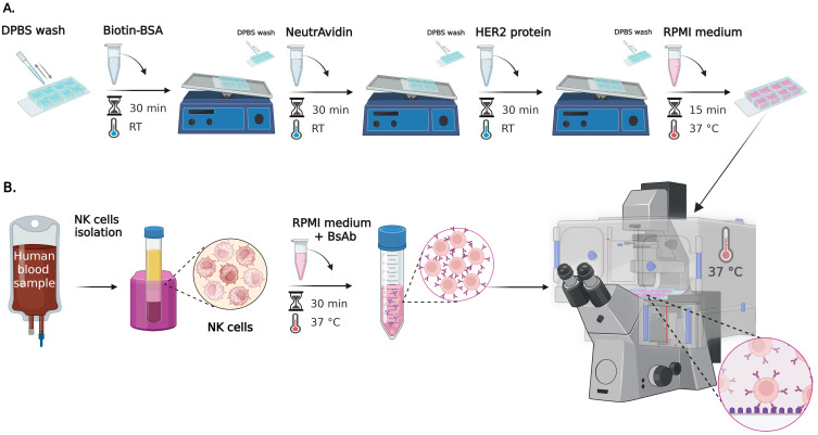
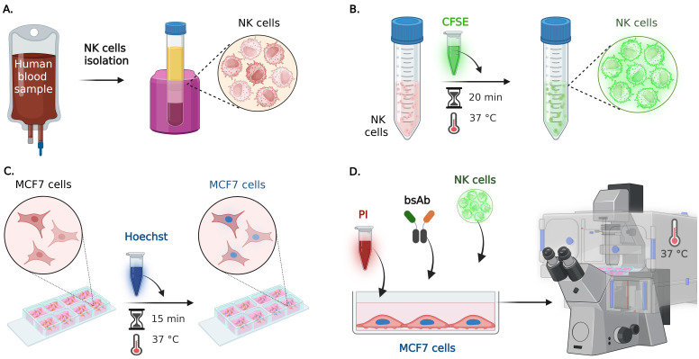
Cell-surface and cell-cell interaction assays are fundamental for studying receptor-ligand interactions and characterizing cellular responses and functions. They play a critical role in diagnostics and in modulating immune system activity for therapeutic applications, notably in cancer immunotherapy. By providing time-lapsed and cell-level direct observation of the sample, optical microscopy offers strong advantages compared to current go-to techniques, which are typically either ensemble methods (e.g., measuring cell populations) or indirect readouts (e.g., impedance for adherent cells). This protocol describes two complementary microscopy-based assays: (1) a cell-surface ligand binding assay to quantify dynamic interactions between human primary Natural Killer (NK) cells and a cancer-mimicking surface, and (2) a cell-cell interaction assay to evaluate antibody-dependent cell cytotoxicity (ADCC) mediated by NK cells targeting tumor cells. Additionally, the protocol uses Celldetective, a new open graphical user interface for quantitative analysis of cell interaction dynamics from 2D time-lapse microscopy datasets. Although applied here to primary immune cells, these methods are adaptable to various cell types, including other immune cells, fibroblasts, and cancer cells. This approach enables direct observation and quantification of cellular morphology, motility, cell-cell interactions, and dynamic behaviors at single-cell resolution over time, facilitating detailed analysis of mechanisms such as cell death, migration, and immune synapse formation. Key features • End-to-end protocol for antibody evaluation by optical microscopy on living cells using simple reagents, followed by full open-source software image analysis and data rendering • Quantitative analysis of cell-surface interactions using label-free imaging to study the dynamic spreading of NK cells on antibody-coated surfaces under different antibody concentrations. • High-resolution evaluation of antibody-dependent cell cytotoxicity in NK-cancer cells co-culture using fluorescence imaging, deep learning-based death detection, and synchronized single-cell measurements.

 求助内容:
求助内容: 应助结果提醒方式:


