{"title":"Trade-Off Analysis of Classical Machine Learning and Deep Learning Models for Robust Brain Tumor Detection: Benchmark Study.","authors":"Yuting Tian","doi":"10.2196/76344","DOIUrl":null,"url":null,"abstract":"<p><strong>Background: </strong>Medical image analysis plays a critical role in brain tumor detection, but training deep learning models often requires large, labeled datasets, which can be time-consuming and costly. This study explores a comparative analysis of machine learning and deep learning models for brain tumor classification, focusing on whether deep learning models are necessary for small medical datasets and whether self-supervised learning can reduce annotation costs.</p><p><strong>Objective: </strong>The primary goal is to evaluate trade-offs between traditional machine learning and deep learning, including self-supervised models under small medical image data. The secondary goal is to assess model robustness, transferability, and generalization through evaluation of unseen data within- and cross-domains.</p><p><strong>Methods: </strong>Four models were compared: (1) support vector machine (SVM) with histogram of oriented gradients (HOG) features, (2) a convolutional neural network based on ResNet18, (3) a transformer-based model using vision transformer (ViT-B/16), and (4) a self-supervised learning approach using Simple Contrastive Learning of Visual Representations (SimCLR). These models were selected to represent diverse paradigms. SVM+HOG represents traditional feature engineering with low computational cost, ResNet18 serves as a well-established convolutional neural network with strong baseline performance, ViT-B/16 leverages self-attention to capture long-range spatial features, and SimCLR enables learning from unlabeled data, potentially reducing annotation costs. The primary dataset consisted of 2870 brain magnetic resonance images across 4 classes: glioma, meningioma, pituitary, and nontumor. All models were trained under consistent settings, including data augmentation, early stopping, and 3 independent runs using the different random seeds to account for performance variability. Performance metrics included accuracy, precision, recall, F<sub>1</sub>-score, and convergence. To assess robustness and generalization capability, evaluation was performed on unseen test data from both the primary and cross datasets. No retraining or test augmentations were applied to the external data, thereby reflecting realistic deployment conditions. The models demonstrated consistently strong performance in both within-domain and cross-domain evaluations.</p><p><strong>Results: </strong>The results revealed distinct trade-offs; ResNet18 achieved the highest validation accuracy (mean 99.77%, SD 0.00%) and the lowest validation loss, along with a weighted test accuracy of 99% within-domain and 95% cross-domain. SimCLR reached a mean validation accuracy of 97.29% (SD 0.86%) and achieved up to 97% weighted test accuracy within-domain and 91% cross-domain, despite requiring 2-stage training phases involving contrastive pretraining followed by linear evaluation. ViT-B/16 reached a mean validation accuracy of 97.36% (SD 0.11%), with a weighted test accuracy of 98% within-domain and 93% cross-domain. SVM+HOG maintained a competitive validation accuracy of 96.51%, with 97% within-domain test accuracy, though its accuracy dropped to 80% cross-domain.</p><p><strong>Conclusions: </strong>The study reveals meaningful trade-offs between model complexity, annotation requirements, and deployment feasibility-critical factors for selecting models in real-world medical imaging applications.</p>","PeriodicalId":73551,"journal":{"name":"JMIR AI","volume":"4 ","pages":"e76344"},"PeriodicalIF":2.0000,"publicationDate":"2025-09-15","publicationTypes":"Journal Article","fieldsOfStudy":null,"isOpenAccess":false,"openAccessPdf":"https://www.ncbi.nlm.nih.gov/pmc/articles/PMC12456844/pdf/","citationCount":"0","resultStr":null,"platform":"Semanticscholar","paperid":null,"PeriodicalName":"JMIR AI","FirstCategoryId":"1085","ListUrlMain":"https://doi.org/10.2196/76344","RegionNum":0,"RegionCategory":null,"ArticlePicture":[],"TitleCN":null,"AbstractTextCN":null,"PMCID":null,"EPubDate":"","PubModel":"","JCR":"","JCRName":"","Score":null,"Total":0}
引用次数: 0
Abstract
Background: Medical image analysis plays a critical role in brain tumor detection, but training deep learning models often requires large, labeled datasets, which can be time-consuming and costly. This study explores a comparative analysis of machine learning and deep learning models for brain tumor classification, focusing on whether deep learning models are necessary for small medical datasets and whether self-supervised learning can reduce annotation costs.
Objective: The primary goal is to evaluate trade-offs between traditional machine learning and deep learning, including self-supervised models under small medical image data. The secondary goal is to assess model robustness, transferability, and generalization through evaluation of unseen data within- and cross-domains.
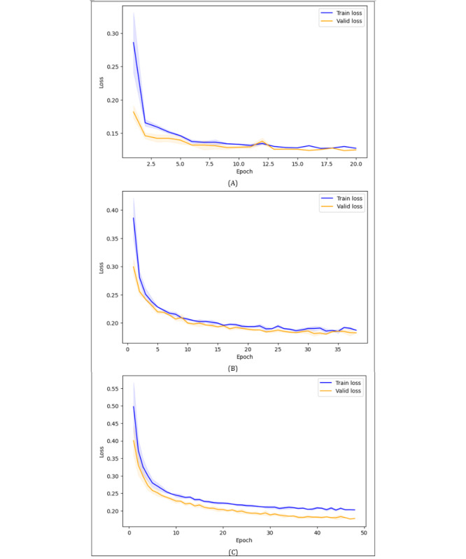
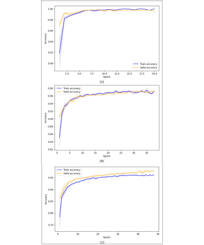
Methods: Four models were compared: (1) support vector machine (SVM) with histogram of oriented gradients (HOG) features, (2) a convolutional neural network based on ResNet18, (3) a transformer-based model using vision transformer (ViT-B/16), and (4) a self-supervised learning approach using Simple Contrastive Learning of Visual Representations (SimCLR). These models were selected to represent diverse paradigms. SVM+HOG represents traditional feature engineering with low computational cost, ResNet18 serves as a well-established convolutional neural network with strong baseline performance, ViT-B/16 leverages self-attention to capture long-range spatial features, and SimCLR enables learning from unlabeled data, potentially reducing annotation costs. The primary dataset consisted of 2870 brain magnetic resonance images across 4 classes: glioma, meningioma, pituitary, and nontumor. All models were trained under consistent settings, including data augmentation, early stopping, and 3 independent runs using the different random seeds to account for performance variability. Performance metrics included accuracy, precision, recall, F1-score, and convergence. To assess robustness and generalization capability, evaluation was performed on unseen test data from both the primary and cross datasets. No retraining or test augmentations were applied to the external data, thereby reflecting realistic deployment conditions. The models demonstrated consistently strong performance in both within-domain and cross-domain evaluations.
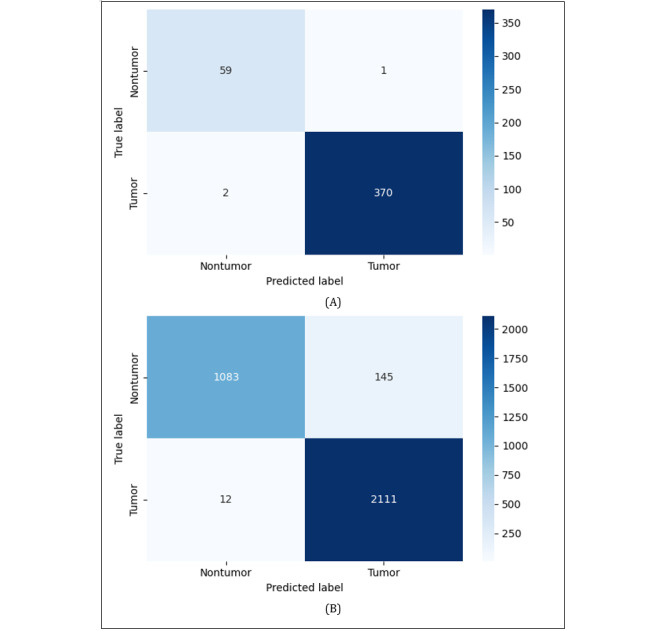
Results: The results revealed distinct trade-offs; ResNet18 achieved the highest validation accuracy (mean 99.77%, SD 0.00%) and the lowest validation loss, along with a weighted test accuracy of 99% within-domain and 95% cross-domain. SimCLR reached a mean validation accuracy of 97.29% (SD 0.86%) and achieved up to 97% weighted test accuracy within-domain and 91% cross-domain, despite requiring 2-stage training phases involving contrastive pretraining followed by linear evaluation. ViT-B/16 reached a mean validation accuracy of 97.36% (SD 0.11%), with a weighted test accuracy of 98% within-domain and 93% cross-domain. SVM+HOG maintained a competitive validation accuracy of 96.51%, with 97% within-domain test accuracy, though its accuracy dropped to 80% cross-domain.
Conclusions: The study reveals meaningful trade-offs between model complexity, annotation requirements, and deployment feasibility-critical factors for selecting models in real-world medical imaging applications.

 求助内容:
求助内容: 应助结果提醒方式:


