An All-Female Study Quantifying Morphological Differences of the Hindfoot and Posterior Tibialis Muscle Between Patients with Progressive Collapsing Foot Deformity and Asymptomatic Controls.
{"title":"An All-Female Study Quantifying Morphological Differences of the Hindfoot and Posterior Tibialis Muscle Between Patients with Progressive Collapsing Foot Deformity and Asymptomatic Controls.","authors":"Takuma Miyamoto, Rich J Lisonbee, Hiroaki Kurokawa, Akira Taniguchi, Yasuhito Tanaka, Amy L Lenz","doi":"10.2106/JBJS.OA.24.00215","DOIUrl":null,"url":null,"abstract":"<p><strong>Background: </strong>The posterior tibialis (PT) muscle and tendon could be a contributing factor in progressive collapsing foot deformity (PCFD). Still, the extent to which the function of the PT is related to PCFD progression is debated. Recently, statistical shape models (SSM) have been shown to provide more accurate and comprehensive morphological evaluations of musculoskeletal tissue. To enhance understanding of the PT in patients with PCFD, we used a 3D SSM to test the hypothesis that both the shape of the PT and alignment of the PT relative to the hindfoot bones are different in female patients with PCFD when compared with female asymptomatic controls.</p><p><strong>Methods: </strong>In this retrospective comparative study, 12 female patients presenting with PCFD and 19 female asymptomatic individuals were included. Computed tomography images were segmented to create 3D models of hindfoot bones and PT. We compared the shape of the PT and alignment of the PT with the hindfoot bones using SSM. The PT 3D model was also used to measure volume, tendon normalized length, and percentage fat of the PT muscle. Each measurement and SSM were compared between PCFD and asymptomatic controls.</p><p><strong>Results: </strong>SSM did not identify significant differences in the isolated shape of the PT between groups. However, the SSM showed significant differences in the alignment of PT tendon regarding the hindfoot bones. Tendon normalized length was significantly lower in PCFD than that in controls. Percent fat content was significantly higher in PCFD compared with controls. No significant differences were found for PT volume between both groups.</p><p><strong>Conclusion: </strong>Our results indicate that in female patients with PCFD, the PT does not differ significantly in shape and volume, only in tendon alignment relative to hindfoot bones, suggesting that the PT may be not always directly involved in the progression of PCFD.</p><p><strong>Clinical relevance: </strong>The application of SSM to assess bones and muscles simultaneously in female patients with PCFD is a new and novel approach to understanding the pathophysiology of this disease.</p><p><strong>Evidence level: </strong>Level III. See Instructions for Authors for a complete description of levels of evidence.</p>","PeriodicalId":36492,"journal":{"name":"JBJS Open Access","volume":"10 3","pages":""},"PeriodicalIF":3.8000,"publicationDate":"2025-09-17","publicationTypes":"Journal Article","fieldsOfStudy":null,"isOpenAccess":false,"openAccessPdf":"https://www.ncbi.nlm.nih.gov/pmc/articles/PMC12431736/pdf/","citationCount":"0","resultStr":null,"platform":"Semanticscholar","paperid":null,"PeriodicalName":"JBJS Open Access","FirstCategoryId":"1085","ListUrlMain":"https://doi.org/10.2106/JBJS.OA.24.00215","RegionNum":0,"RegionCategory":null,"ArticlePicture":[],"TitleCN":null,"AbstractTextCN":null,"PMCID":null,"EPubDate":"2025/7/1 0:00:00","PubModel":"eCollection","JCR":"Q2","JCRName":"ORTHOPEDICS","Score":null,"Total":0}
引用次数: 0
Abstract
Background: The posterior tibialis (PT) muscle and tendon could be a contributing factor in progressive collapsing foot deformity (PCFD). Still, the extent to which the function of the PT is related to PCFD progression is debated. Recently, statistical shape models (SSM) have been shown to provide more accurate and comprehensive morphological evaluations of musculoskeletal tissue. To enhance understanding of the PT in patients with PCFD, we used a 3D SSM to test the hypothesis that both the shape of the PT and alignment of the PT relative to the hindfoot bones are different in female patients with PCFD when compared with female asymptomatic controls.
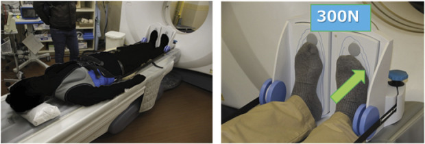
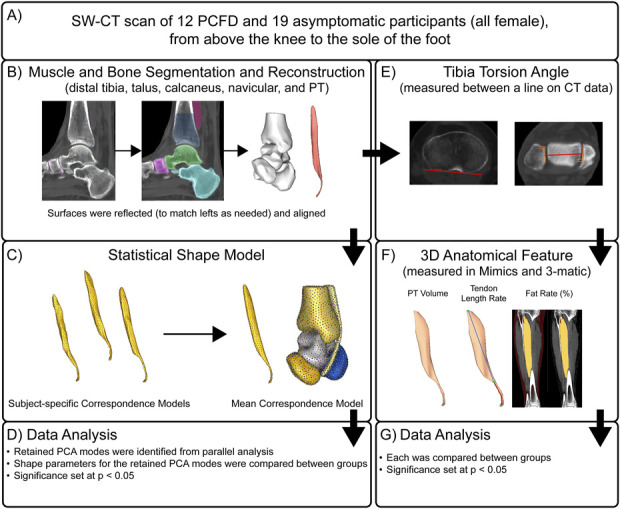
Methods: In this retrospective comparative study, 12 female patients presenting with PCFD and 19 female asymptomatic individuals were included. Computed tomography images were segmented to create 3D models of hindfoot bones and PT. We compared the shape of the PT and alignment of the PT with the hindfoot bones using SSM. The PT 3D model was also used to measure volume, tendon normalized length, and percentage fat of the PT muscle. Each measurement and SSM were compared between PCFD and asymptomatic controls.
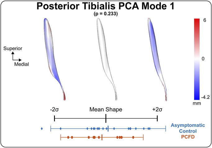
Results: SSM did not identify significant differences in the isolated shape of the PT between groups. However, the SSM showed significant differences in the alignment of PT tendon regarding the hindfoot bones. Tendon normalized length was significantly lower in PCFD than that in controls. Percent fat content was significantly higher in PCFD compared with controls. No significant differences were found for PT volume between both groups.
Conclusion: Our results indicate that in female patients with PCFD, the PT does not differ significantly in shape and volume, only in tendon alignment relative to hindfoot bones, suggesting that the PT may be not always directly involved in the progression of PCFD.
Clinical relevance: The application of SSM to assess bones and muscles simultaneously in female patients with PCFD is a new and novel approach to understanding the pathophysiology of this disease.
Evidence level: Level III. See Instructions for Authors for a complete description of levels of evidence.

 求助内容:
求助内容: 应助结果提醒方式:


